Valvular heart diseases are well studied and there are many ways to carry out treatment, but the moment the disease is discovered is still of great importance.
When the diagnosis arrives at a later stage, treatment will be more difficult and will likely require heart valve surgery.
The earlier the problem is detected and under the control of the doctor, the more favorable the outcome will be for the patients.

What are heart valves?
Heart valves are anatomical structures whose function is extremely important for the health and function of the heart. They are responsible for ensuring that blood flows in one direction through the heart chambers, as well as for the one-way flow of blood from the heart to the large blood vessels.
There are four heart valves, two located on the left side (mitral and aortic), and two on the right (tricuspid and pulmonary).
- Mitral valve (bicuspid valve) – this valve is located between the left atrium and ventricle, and consists of two leaflets (anterior and posterior).
- Tricuspid valve – this valve is located between the right atrium and ventricle, and consists of three leaflets (anterior, posterior, and septal).
- Aortic valve – located between the left ventricle and aorta, it allows for one-way flow of blood from the ventricle to the aorta.
- Pulmonary valve – located between the right ventricle and pulmonary artery, it allows for one-way flow of blood from the heart to the pulmonary artery.
Tricuspid and mitral valves are collectively referred to as atrioventricular (AV) valves, as they are attached to the fibrous skeleton of the heart and separate the heart chambers from the atria, and also allow for one-way blood flow from the atria to the ventricles.
The semilunar valves are the aortic and pulmonary valves.
Heart valve and heart murmur
A heart murmur is an extremely common occurrence, especially in children. In very few cases, it is a reason for concern and further testing. In children, it is usually the result of a congenital heart defect, while in adults, it reflects a problem with the heart, which can be congenital or acquired.
Heart valves, whose main function is to allow blood to flow in one direction, can be the cause of a heart murmur.
If we look at the phase of the cardiac cycle in which the murmur occurs, it can be systolic or diastolic. Systolic murmur is usually benign, or physiological, while diastolic is pathological, or a problem and a reason for treatment.
A heart murmur is easy to detect. A general practitioner can hear it during a simple examination with a stethoscope, and in most cases, this is how it happens, and the doctor then refers the patient to a cardiologist.
What heart valve diseases exist?
- Aortic stenosis
This represents a disease of the aortic valve that involves the narrowing of the aortic opening, causing problems with blood flow from the left ventricle into the aorta. This disease can be congenital or acquired.
If it’s congenital, patients typically have more or fewer leaflets in the valve than they should. The first symptoms usually don’t appear immediately after birth, or even in childhood, but later in the third decade of life.
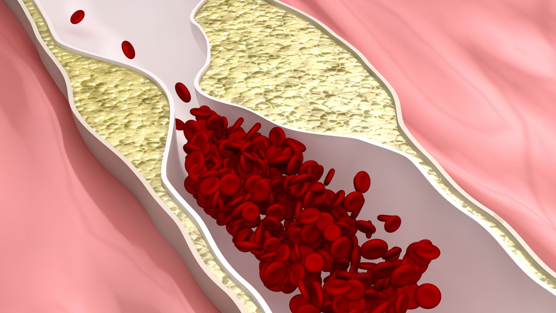
Acquired aortic stenosis can result from rheumatic fever or atherosclerosis. In fact, in the last few decades, the latter has been present in 90% of people with aortic stenosis.
Symptoms often do not occur with this disease, as with most other cardiovascular diseases, especially in the early stages. Sometimes it is discovered accidentally during routine check-ups, but when it does cause symptoms, it has already progressed and burdened the heart significantly. If left untreated, this disease leads to fatal outcomes in symptomatic patients. Within the first five years, 50% of symptomatic patients die, and within ten years, 90%. Patients who undergo treatment have much better chances of a normal and quality life, as the treatment methods are reliable and effective.
- Aortic insufficiency
This type of disease involves a problem with the basic function of the valve, which is to allow one-way blood flow, in this case from the left ventricle to the aorta. In patients with aortic insufficiency, the valve does not function properly, so blood returns from the aorta to the chamber.
This disease can be acute and chronic. Acute develops suddenly and progresses quickly, while in chronic cases, symptoms slowly develop and progress.
The success of treatment depends on the phase in which the patient is. When it comes to patients with a mild or moderate presentation, the survival rate is 80-90%. Even when a person has a severe form of this disease, if valve surgery is performed in time, the prognosis is good.
- Mitral insufficiency
In the case of this disease, there is a backflow of blood from the left ventricle to the left atrium during systole.
The reasons for the appearance of this disease can be mitral valve prolapse and problems with the function of the papillary muscles due to ischemic disease.
Often, years pass before the first symptoms of mitral valve problems appear.
- Mitral stenosis
The nature of this disease is associated with rheumatic endocarditis and it is very rarely hereditary. It involves a narrowing of the opening between the left atrium and left ventricle, which makes blood flow difficult.
- Tricuspid insufficiency
Tricuspid insufficiency is characterized by insufficient closure, causing blood to flow back from the right ventricle to the right atrium. It most commonly occurs as a result of pulmonary hypertension, infectious endocarditis, rheumatic endocarditis, tumors, and poor papillary muscle function due to ischemic disease.
What are the symptoms of heart valve disease?
Heart valve disease may not show any alarming symptoms immediately. Sometimes, it can take up to ten years before the first discomfort or feeling that something is wrong occurs.
People who develop problems with one or more valves may experience:
- Fatigue
- Weakness
- Heart palpitations
- Shortness of breath
- Coughing or wheezing
- Dizziness
- Fainting
- Chest pain or discomfort
- Swelling in the ankles or feet
- Discomfort when lying down
Symptoms that suggest the disease has progressed may include:
- Loss of consciousness
- Severe, angina-like chest pain
- Fatigue and shortness of breath when lying down
How to diagnose heart valve diseases?
The diagnosis proceeds in the following steps:
- Clinical examination and medical history taking – the patient should describe their symptoms and when they occur in detail.
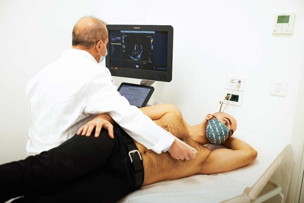
- Echocardiography (ECHO ) ultrasound of the heart – a non-invasive and highly precise method of imaging the heart, essential in this situation as it provides insight into the anatomy of the heart.
- Electrocardiography (ECG) – a non-invasive method of recording the electrical activity of the heart.
Treatment of heart valve diseases
Depending on the patient’s age, condition, and the phase of the disease, a cardiology specialist will recommend the most suitable method of treatment.
The first method involves using medications, whose effect unfortunately does not prevent the progression of the disease, but affects the subjective feeling and symptoms.
The second method is heart valve surgery.
Heart valve surgery is a minimally invasive procedure. Again, depending on the need, valve reconstruction or replacement is performed. In patients at risk for heart valve surgery, transcatheter implantation is carried out. The most common is surgical intervention to replace the aortic valve.
The prosthesis that is placed instead of the natural valve can be mechanical or biological. The biological prosthesis is made from a pig or cow valve and has a lifespan of about 10 years, which in most cases means that the patient will need further replacement surgeries. The mechanical prosthesis lasts a lifetime, but also involves regularly taking blood-thinning medication. When a biological prosthesis is implanted, blood-thinning medications are used for a shorter period.
Each prosthesis has its advantages and situations in which they are better. Reconstruction of the valve is most commonly performed on the mitral valve. What is an advantage here is the preservation of the valve, so blood-thinning medications are not necessary for the patient. If you have any doubts about these conditions and are interested in which treatment method is best and safest for you, schedule an appointment with one of our cardiologists.