Home » Services » Ultrasound (Ultrasound Diagnostics) » Ultrasound Examination of the Thyroid Gland
Ultrasound Examination of the Thyroid Gland
What is a thyroid ultrasound?
Neck and thyroid ultrasound is a diagnostic procedure used to assess the condition of the thyroid gland and surrounding structures in the neck. Thyroid ultrasound is a non-invasive medical procedure used to visualize and evaluate the condition of the thyroid gland using ultrasound waves. The thyroid gland is a small gland located in the front of the neck, under the “Adam’s apple”, and plays a key role in the regulation of the body’s metabolism through the production of thyroid hormones (thyroxine – T4 and triiodothyronine – T3).
Thyroid ultrasound is a painless, quick and safe procedure. There is no exposure to ionizing radiation, which makes it safe and suitable for repeated examinations. The results of an ultrasound examination often help doctors make an accurate diagnosis and plan further treatment, if necessary.
Why is an ultrasound examination of the thyroid gland being performed?
- Thyroid examination includes: medical history, physical examination of the thyroid gland, blood laboratory parameters and ultrasound of the thyroid gland.
- In the event of the appearance of symptoms that may indicate problems with the thyroid gland: weight changes, fatigue and exhaustion, insomnia or sleep disorders, mood changes, fast or slow heart rate, increased blood pressure or palpitations, irregular or heavy periods, dry skin, brittle hair, hair loss, neck swelling
- Due to disturbed values of laboratory parameters, imaging of the thyroid gland is performed: thyroid hormones, triiodothyronine (T3) and thyroxine (T4), thyrotropin hormone (TSH), antibodies to the thyroid gland
- Thyroid ultrasound is performed if the thyroid gland is overactive when the following symptoms occur: increased appetite, weight loss, increased irritability, nervousness and restlessness, rapid heartbeat, sweating and sensitivity to heat, changes in the menstrual cycle in women, weak tolerance to physical exertion, restless sleep changes in the eyes, such as bulging eyes (exophthalmos) and dry eye.
- Thyroid ultrasound and hormones: two important diagnostic procedures used to assess thyroid health and function. If TSH is increased and T4 and T3 are decreased, it may indicate hypothyroidism (decreased function). If TSH is decreased and T3 and T4 are elevated, it may indicate hyperthyroidism (increased function).
- The presence of thyroid antibodies (anti TPO At, anti TG At): may indicate autoimmune diseases of the thyroid gland, such as Hashimoto’s thyroiditis or Graves’ disease
- Detection of nodules and other changes: Ultrasound allows the identification and evaluation of nodules, cysts or other unusual formations in the thyroid gland. These changes can be benign or malignant, so ultrasound is used for their early detection and risk assessment.
- Follow-up of thyroid disease: Patients with diagnosed thyroid disease, such as hypothyroidism, hyperthyroidism, or autoimmune diseases, undergo regular ultrasound examinations to monitor the progress of the disease, the effectiveness of therapy, and the eventual appearance of new problems.
- Assessment of thyroid function: Ultrasound can reveal signs of inflammation (thyroiditis) or other structural changes that may affect thyroid function. This helps doctors better understand the condition of the gland and adjust therapy if necessary.
- Follow-up of previously identified nodules: If previous examinations, such as palpation of the neck or other diagnostic methods, have revealed nodules in the thyroid gland, ultrasound can provide additional information about their size, shape and structure.
- Thyroid cancer risk assessment: Ultrasound can help assess the likelihood that malignant changes are present in the thyroid gland, allowing doctors to make decisions about further diagnostic tests or a biopsy if needed.
- Follow-up after surgery: Patients who have previously undergone surgery on the thyroid gland should be regularly monitored with ultrasound to assess the possible return of problems or complications after surgery.
Ultrasound examination of the thyroid gland is a painless, quick and safe procedure that enables a detailed examination of this important organ without exposing the patient to ionizing radiation. The results of this examination can be of crucial importance for the diagnosis, treatment and monitoring of the condition of the thyroid gland.
What does a thyroid ultrasound show?
Size and shape
Nodules and changes
Cysts
Diffuse changes
Blood vessels
Goiter
Parathyroid glands
How to prepare for thyroid ultrasound?
How is thyroid ultrasound performed?
- Patient preparation: The patient should be placed in a supine or sitting position with the neck slightly tilted back to facilitate access to the front of the neck.
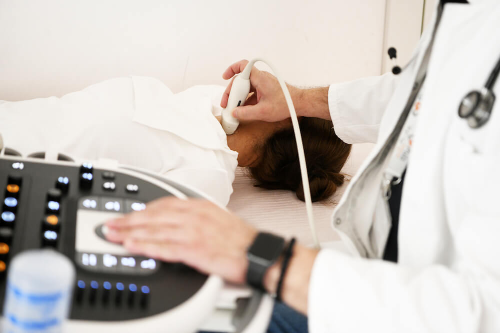
- Ultrasound gel: Before the examination begins, the medical technician (sonographer) will apply gel to the front of the neck. The gel helps ensure good contact between the ultrasound probe and the skin, which improves image quality.
- Using the ultrasound probe: The sonographer will hold an ultrasound probe (which is similar to a small microphone) to the front of the neck. The probe emits high-frequency sound waves into the body and then registers the reflected waves that return. This information is converted into an image of the thyroid gland on the screen of the ultrasound machine.
- Probe movement: The sonographer will carefully move the probe across the front of the neck to examine the entire thyroid gland and surrounding structures. During the examination, the sonographer may ask the patient to change the position of the head or take a deep breath to better visualize certain regions.
- Imaging and measurement: During the examination, the sonographer may record images of the thyroid gland and measure the dimensions of nodules or other changes that are detected.
- Completion of the examination: When the examination is complete, the sonographer will wipe the gel from the patient’s neck.
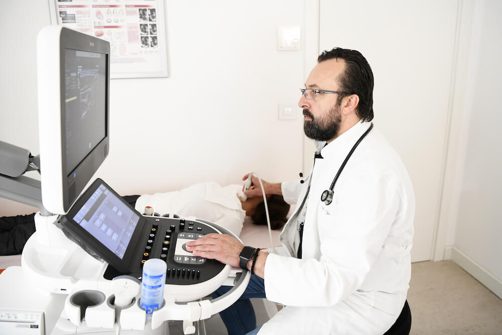
What does ultrasound equipment look like?
Ultrasound machine
Ultrasound probe
Monitor
Control keyboard
Printer (optional)
Ultrasound gel
What can I expect during and after the thyroid gland ultrasound examination?
- Preparing for the examination: Before the actual examination, you may be asked to change into a hospital gown or to remove necklaces and other jewellery that may obstruct access to the front of the neck.
- Examination position: You will be placed in a lying or sitting position with your neck slightly tilted back so that the sonographer can access the front of your neck.
- Applying the gel: The sonographer will apply the gel to the front of the neck. This gel helps ensure good contact between the ultrasound probe and the skin, which improves image quality.
- Ultrasound examination: The sonographer will carefully hold the ultrasound probe at the front of the neck and move it to examine the entire thyroid gland and surrounding structures. This procedure is painless and usually does not cause discomfort.
- 5. Analysis of the results: The results of the ultrasound of the thyroid gland are immediately displayed on the screen. The sonographer will analyze the images and measure any changes detected during the examination.
- Gel removal: After the examination is complete, the sonographer will wipe the remaining gel from the front of the neck.
- Return to normal activities: Thyroid ultrasound does not require recovery and you can immediately return to your normal activities.
- Interpretation of the results: The results of the examination will be available to the doctor who will analyze and interpret them. Your doctor will explain the findings and, if necessary, take additional steps regarding further diagnosis or treatment.
How long does a thyroid ultrasound exam take?
Who interprets thyroid ultrasound results and how do I get them?
What are the risks of thyroid ultrasound examination?
What are the benefits of thyroid ultrasound examination?
- Safety: Ultrasound is a safe examination because it does not use ionizing radiation, unlike some other diagnostic methods such as X-rays and CT scans. This means that there is no risk of exposing the patient to harmful radiation.
- Painlessness: Ultrasound is a non-invasive and usually painless procedure. No need to prick the skin or use a needle for this test. The probe is simply held on the surface of the skin to examine the inside of the neck.
- Speed: An ultrasound examination of the thyroid gland is a quick procedure that usually takes only a few minutes. The results are available immediately and can be used for rapid diagnosis and planning of further treatment steps, if necessary.
- Accuracy: Ultrasound of the thyroid gland provides detailed images of the inside of the gland, which allows for a precise analysis of its dimensions, shape, structure and possible changes.
- Monitoring over time: Ultrasound can be useful for monitoring the condition of the thyroid gland over time. If changes are detected, the ultrasound can be repeated to monitor their behavior and possibly respond to them.
- Aid in diagnosis: Ultrasound can help in the diagnosis of various thyroid conditions, including the presence of nodules, cysts, inflammatory or autoimmune changes.
- Safe for pregnant women: Ultrasound is safe to use during pregnancy, making it useful for monitoring thyroid conditions in pregnant women.
What are the limitations of thyroid ultrasound?
It cannot differentiate between benign and malignant diseases:
Nodules that are not big enough:
View limited to the surface
Does not detect every type of thyroid disease
The doctor may indicate further diagnostic methods such as:
- Scintigraphy: Thyroid scintigraphy is a nuclear medicine procedure that uses radioactive substances (usually iodine) to show the functionality of the thyroid gland. This method can help identify hot or cold nodes, which indicate certain functional disorders.
- Fine needle biopsy (fine needle biopsies): If thyroid nodules are found, the doctor may decide to perform a fine needle biopsy. This procedure involves taking a tissue sample from the nodule for further analysis under a microscope to determine whether the change is benign or malignant.
- Thyroid calcium scan: This procedure can be useful for evaluating the presence of calcifications in the thyroid gland, which may be associated with certain diseases.
- Other diagnostic methods: In some cases, the doctor may order additional tests and analyses, such as a CT scan, magnetic resonance imaging (MRI) or other nuclear medicine procedures, in order to better evaluate the thyroid gland and any changes.
Ultrasound of the thyroid gland – Price
Price of this exam is 5000 RSD.