Home » Services » Ultrasound (Ultrasound Diagnostics) » Doppler of Abdominal Aorta With Visceral Branches
Doppler of abdominal aorta with visceral branches
Anatomy of the Abdominal Aorta
The aorta is the largest artery in the human body and plays a key role in blood circulation. It begins as the ascending part of the aorta (aorta ascendens) from where the blood vessels that supply the heart with oxygen go. It continues with the Aortic arch, from which the branches that supply blood to the head and upper extremities depart. Then continues the descending part of the aorta (aorta desecedens): the aorta descends through the chest (thorax) and is called the thoracic aorta which supplies blood to the organs and structures in the chest and after passing through the diaphragm, the aorta enters the abdominal part of the body and is called the abdominal aorta. Here it divides into different branches that supply blood to the organs in the abdomen and pelvis.
The abdominal aorta is a part of the aorta located in the abdominal part of the body and extends from the diaphragm at the level of the lower edge of the body of the 12th thoracic vertebra to its division into the iliac arteries, at the level of the body of the 4th lumbar vertebra, which supply blood to the pelvis and lower extremities. The abdominal aorta has several important branches that supply various organs and structures in the abdominal cavity. During its journey through the abdomen, it gives off its visceral and parietal branches.
Branches to the abdominal wall:
The abdominal aorta gives off branches that supply blood to the muscles and skin of the abdominal wall. This includes the inferior epigastric artery, which supplies the anterior abdominal wall.
Visceral branches
They serve to nourish the organs of the abdominal cavity, and the most important are:
The celiac trunk (lat. Truncus coeliacis)
It arises from the abdominal aorta as soon as it passes through the diaphragm and is divided into:
- The left gastric artery (lat. Arteria gastrica sinistra), which supplies blood to the stomach
- Common hepatic artery (lat. Arteria hepatica) from which branches supply the liver, gallbladder and part of the stomach.
- Splenic artery (lat. Arteria lienalis), which supplies blood to the spleen
- Lower pancreatic (lat. Arteria pancreaticoduodenalis inferior) which supplies blood to the pancreas
The superior mesenteric artery (lat. Arteria mesenterica superior)
It is responsible for supplying blood to most of the small intestine (jejunum and ileum), the upper part of the large intestine (including the appendix and the part of the large intestine called the transverse colon), and the pancreas (pancreas).
The left and right renal arteries (lat. Arteria renalis sinistra et dextra)
Those are arteries that supply blood to the kidneys. Each renal artery enters the kidney and further branches into smaller arteries and arterioles that penetrate the renal tissue.
The lower mesenteric artery (lat. Arteria mesenterica inferior)
It is an artery that separates from the abdominal aorta and is responsible for supplying blood to the lower part of the large intestine (sigmoid colon and rectum), as well as the parts of the large intestine above the rectum.
Abdominal aorta and its visceral branches are of normal size
Measurements of the abdominal aorta are often taken to assess the health of this vital blood vessel and identify possible problems, such as an aneurysm (widening of the artery wall). These measurements are usually expressed in millimetres (mm) and take into account the width (diameter) of the aorta. Normal measurements of the abdominal aorta vary depending on age, gender, and other factors, but it is generally considered that the normal diameter of the abdominal aorta is less than 3 cm to 3.5 cm.
Measuring the dimensions of the visceral branches of the abdominal aorta can be part of the diagnostic process when problems or diseases are suspected in the organs supplied with blood by these arteries. The size of the diameter of the arteries can vary between individuals and depending on various factors, including anatomical variability and individual characteristics.
Normal diameter of visceral branches
Truncus coeliacus: It is considered that the normal diameter of this artery is in the normal range between 7 and 15 millimetres (0.7 – 1.5 centimetres). This size range is usually considered normal and does not indicate a narrowing (stenosis) or enlargement of the artery.
Arteria hepatica (liver artery): The normal diameter of the hepatic artery propria, the main branch of the hepatic artery, is usually between 3 and 5 millimetres (0.3 – 0.5 centimetres). The diameter can be slightly larger when this artery branches into smaller branches that supply different parts of the liver.
Arteria gastrica (gastric artery): The normal diameter of arteria gastrica sinistra (left coronary artery) and arteria gastrica dextra (right coronary artery) can range from 1 to 2 millimeters (0.1 – 0.2 centimetres) each.
Arteria lienalis (splenic artery): The normal diameter of the arteria lienalis usually ranges between 1 and 2 millimetres (0.1 – 0.2 centimetres).
The superior mesenteric artery ranges between 6 and 10 millimetres (0.6 – 1.0 centimetres). This range is considered normal and usually does not indicate narrowing (stenosis) or enlargement of the artery.
The inferior mesenteric artery normally ranges between 5 and 10 millimetres (0.5 – 1.0 centimetres). This size range is usually considered normal and does not indicate a narrowing (stenosis) or enlargement of the artery.
Arteria renalis (renal arteries): The normal diameter can vary among individuals, but the diameter of these arteries is usually considered to be in the normal range between 4 and 6 millimetres (0.4 – 0.6 centimetres).
What is abdominal aortic doppler?
Abdominal aortic Doppler is a medical procedure that uses the Doppler effect to evaluate blood flow through the abdominal aorta and identify potential problems, such as narrowings (stenosis), aneurysms (dilations), or other vascular disorders.
The Doppler effect is used so that high-frequency sound waves are emitted towards the abdominal aorta, and then their reflection from the blood flow inside the artery is detected. Based on changes in the frequency of these reflections (the so-called Doppler effect), the doctor can determine the speed and direction of the blood in the aorta. This allows assessment of blood flow through the aorta and identification of any abnormalities.
This procedure is non-invasive, meaning no surgery is required, and is usually performed using an ultrasound machine. Abdominal aortic Doppler results can help doctors make decisions about further treatment or monitoring of the patient’s condition.
Who should do abdominal aortic doppler and when?
This test is usually ordered when problems with the abdominal aorta or other large arteries in the abdominal cavity are suspected.
Here are some situations in which Doppler of the abdominal aorta may be recommended:
- Patients with symptoms or risk factors: People who have symptoms suggestive of a vascular problem in the abdomen, such as abdominal pain or discomfort, a pulsating tumor in the abdomen, or back pain, may be candidates for this procedure. Also, patients with risk factors, such as smoking, high blood pressure, diabetes, or a family history of vascular disease, may be referred for abdominal aortic Doppler to assess the condition.
- Patients with a history of aortic problems: People with a history of aortic disease, such as aortic aneurysm or aortic atherosclerosis, are often followed through regular Doppler examinations to monitor the condition and identify changes over time.
- Routine screening tests: In some cases, the doctor may recommend a Doppler of the abdominal aorta as part of a routine examination for certain categories of patients, especially if there are risk factors.
- Patients who are preparing for surgery or an invasive procedure: Before a planned surgical intervention in the abdomen, the doctor can order a Doppler of the abdominal aorta to assess the patency of the arteries and identify possible problems that could affect the operation.
What are the common pathological conditions that require Doppler?
Atherosclerosis of the abdominal aorta is a condition in which plaques, consisting of fatty deposits, cholesterol, calcium and other materials, build up inside the walls of the abdominal aorta. These plaque buildups can cause narrowing (stenosis) or blockage of the artery, which can lead to serious complications, such as ischemia of the abdominal organs or an increased risk of aortic aneurysm (enlargement of the aorta).
Anomalies and variations of the abdominal aorta
The abdominal aorta can have various anomalies and variations in its anatomical shape and structure. Most of these variations are not associated with significant health problems and can be present without causing symptoms or complications. However, some of these anomalies can have a potential impact on health, especially if complications occur. There may be an abnormal position of the abdominal aorta which is usually not a problem if it does not interfere with normal blood flow or press on surrounding structures. Coarctation of the abdominal aorta is a narrowing of the abdominal aorta that can restrict blood flow to the lower body. It can be present from birth (congenital coarctation) or it can develop later in life. The abdominal aorta usually branches into several smaller arteries that supply the organs in the abdominal cavity. Variations in the number or arrangement of these branches may exist, but usually do not cause problems unless they cause troublesome vascular anomalies.
Changes in the size of the abdominal aorta can be a sign of different conditions and can have different health consequences. Here are some possible scenarios of changes in the size of the abdominal aorta and their implications:
- Aneurysm of the abdominal aorta: An aneurysm of the abdominal aorta is the enlargement of this blood vessel. An aneurysm is usually considered serious because there is a risk of the aorta rupturing, which can be potentially fatal. Monitoring and treatment of the aneurysm may be necessary to prevent complications.
- Narrowing of the abdominal aorta (coarticulation): Narrowing of the abdominal aorta can restrict blood flow to the lower body. This condition can cause hypertension (high blood pressure) and other circulation problems. In more severe cases, surgery may be required to correct the problem.
- Variations in the size of the arteries: Normally, the abdominal aorta gradually narrows as it descends towards the lower body. However, there may be variations in the size and shape of the abdominal aorta in some people. These variations usually do not cause problems if they do not interfere with normal blood flow.
- Changes during aging: With aging, the abdominal aorta can become smaller and lose some elasticity. This is a natural process of aging and usually does not cause problems unless the person already has other vascular diseases.
- Tumor or other compressions: The presence of a tumor, cyst or other mass near the abdominal aorta can put pressure on this artery and cause changes in its shape or size. This can lead to circulation problems and should be carefully monitored and treated.
Abdominal aortic aneurysm is a serious condition in which a part of the abdominal aorta becomes enlarged (dilatation), which can pose a serious health risk. These enlargements usually occur in the lower part of the abdominal aorta and can be life-threatening if the aneurysm ruptures.
An abdominal aortic aneurysm usually causes no symptoms as long as it is small and stable. However, when the aneurysm becomes larger or begins to spread, it can cause pain in the lower abdomen or back. If an aneurysm ruptures, it can lead to sudden and severe abdominal pain, shock, and often death.
Risk factors for the existence of an aneurysm
The most common cause of abdominal aortic aneurysm is atherosclerosis, which is the buildup of plaque within the arterial walls. Other risk factors include smoking, high blood pressure, genetic predisposition, male gender (aneurysms are more common in men), high blood pressure, high cholesterol, diabetes, and older age.
Abdominal aortic aneurysm screening is a preventive medical test performed to detect aneurysms in this part of the aorta before they become serious health problems. This screening is usually recommended for people who are at increased risk for developing an aneurysm, especially men over the age of 65 and people who have the risk factors listed above.
The most common screening method for abdominal aortic aneurysm is ultrasound (Doppler ultrasound), which is non-invasive and painless. Ultrasound allows doctors to visualize the aorta and measure its size, identifying possible aneurysms.
If during the screening it is determined that the person has an abdominal aortic aneurysm, the doctor will determine the next steps. This may include regular monitoring of the size of the aneurysm, treatment, or surgery, depending on the size and other factors.
Treatment of abdominal aortic aneurysm
Treatment of aneurysms depends on their size, location, and other factors, and may involve a variety of therapeutic approaches, including:
Monitoring and control of risk factors
If the aneurysm is small and stable, the doctor may recommend regular monitoring of the size of the aneurysm by ultrasound or other diagnostic methods. Also, it is important to control risk factors such as smoking, high blood pressure, and high cholesterol levels to prevent further aneurysm growth.
Surgery
If the aneurysm is large, growing rapidly, or at risk of rupture, surgery may be necessary. There are two main types of surgery to treat aneurysms:
Open abdominal aortic aneurysm surgery: This invasive procedure involves opening the abdominal cavity and replacing the diseased aorta with a prosthesis or patch to ensure normal blood flow. Open surgery is usually necessary for larger aneurysms or those that have ruptured.
Endovascular Abdominal Aortic Aneurysm Surgery: This less invasive procedure involves the use of a catheter that is inserted through the blood vessels to repair the aneurysm. A stent-graft (metal mesh covered with fabric) is placed inside the aneurysm to strengthen the arterial wall and prevent further expansion. Endovascular surgery may be an option for certain patients, but is not always possible, especially if the aneurysm has certain characteristics.
Drug therapy
The doctor may recommend drugs to control risk factors, such as drugs to lower blood pressure, drugs to control cholesterol, and antiplatelet drugs that prevent blood clotting.
Lifestyle changes
People with aneurysms should talk to their doctor about lifestyle changes that can reduce the risk of complications. These may include quitting smoking, eating a healthy diet, exercising regularly and controlling risk factors.
Aneurysm treatment requires careful assessment and an individual approach to each patient. It is important that a person with an aneurysm works with their doctor to make the best treatment decision and follow up with the necessary therapy to prevent complications. Aneurysms are a serious condition and should be treated under the supervision of a vascular specialist.
Why doppler abdominal aorta is needed?
Abdominal aortic Doppler is an important tool for diagnosis, monitoring and treatment planning of vascular problems in this part of the body. It allows accurate and non-invasive examination of blood vessels, which is essential for preserving the health of the heart and blood vessels, and which is often used for several reasons:
Early detection of aneurysms
One of the main reasons for using Doppler of the abdominal aorta is the early detection of aneurysms (dilation) in this part of the aorta. Abdominal aortic aneurysms often have no symptoms when they are small and stable, but they can be serious and potentially fatal if they rupture. Doppler allows doctors to accurately measure the size of the aorta and identify the presence of an aneurysm.
Assessment of blood flow
Doppler ultrasound allows doctors to assess the speed and direction of blood flow through the abdominal aorta. This is useful for identifying possible narrowings (stenosis) or blockages in the artery, which may indicate atherosclerosis or other vascular problems.
Monitoring vascular problems
For people who have already been diagnosed with atherosclerosis or abdominal aortic aneurysm, Doppler is often used to monitor the progress of the disease over time. Regular follow-up can help in making treatment decisions and identifying possible complications.
Treatment planning
If a vascular problem, such as a narrowing or aneurysm, is detected, Doppler can help doctors plan appropriate treatment. Based on the Doppler results, the doctor can make a decision about surgery, endovascular procedure or medical treatment.
Controlling risk factors
Doppler can help doctors identify risk factors for vascular disease, such as narrowing of the arteries or damage to the arterial walls. It can be beneficial for patients to change their lifestyle and reduce the risk of further vascular problems.
When do symptoms affect specific parts of the body?
Disorders of blood vessels in the abdomen can be asymptomatic in the initial stages, so regular monitoring and screening is recommended for people with risk factors. Here are some symptoms that can occur with various disorders of blood vessels in the abdomen:
Abdominal or back pain: Pain can be a symptom of various vascular problems in the abdomen, including aneurysms, narrowed arteries, or thrombosis.
Pulsating abdominal tumor: Abdominal aortic aneurysms can cause a pulsating sensation in the abdomen that may feel like a pulsating abdominal tumor.
Cold extremities: Narrowing or blockage of the arteries in the abdomen can interfere with normal blood flow to the lower extremities, which can lead to cold and pale legs or legs that are often tired or painful when walking (limping).
Abnormal sounds in the abdomen: Sometimes a murmur can be heard in the abdomen (abdominal murmur) which may indicate turbulent blood flow in the arteries.
Weight loss for no apparent reason: In some vascular problems, weight loss can be a symptom, especially if the disorder interferes with normal nutrition and circulation.
Nausea or vomiting: An abdominal aortic aneurysm that is larger or has complications can press on adjacent organs and cause gastrointestinal symptoms such as nausea and vomiting.
Aneurysm Rupture: In the event that an abdominal aortic aneurysm ruptures, you will experience sudden and severe abdominal pain, which may be a symptom of a serious and urgent medical situation.
Gallbladder disorders usually involve problems with gallbladder function or the presence of gallstones. Symptoms can vary depending on the nature of the disorder, but common symptoms include: Pain in the upper right side of the abdomen, pain that may radiate to the right shoulder or back, nausea and vomiting, bloating and gas, heartburn, belching, greasy stools or diarrhea, sometimes yellow discoloration of the skin.
Liver disorders can vary in severity and symptoms, and symptoms are often absent in the early stages of the disease. Symptoms also vary depending on the specific liver disorder. However, here are some common symptoms that may indicate liver disorders: tiredness and exhaustion, pain or discomfort in the upper right side of the abdomen, loss of appetite, nausea and vomiting, darker urine, lighter stools, swelling of the abdomen or legs, yellowing of the skin and whites eyes, itchy skin, weight gain, bleeding disorders, confusion, concentration problems and behavioural changes.
Bowel disorders can include a variety of diseases and conditions that affect the function and structure of the bowel. Symptoms of bowel disorders vary depending on the specific disorder, but common symptoms include: abdominal pain that can occur anywhere along the bowel and may be cramping, dull, sharp or painful stool changes may include diarrhea (frequent, loose stools), constipation (difficulty having a bowel movement), stools that are too greasy or contain blood, mucus or unusual colours, flatulence (flatulence) and frequent gas, nausea and vomiting, blood in the stool, weight loss, tiredness and weakness, changes in appetite, feeling of incomplete emptying of the bowels, altered bowel habits, such as frequent changes between diarrhea and constipation.
Kidney disorders can range from mild to severe, and symptoms vary depending on the type of disorder and the degree of kidney damage. Here are some common symptoms that often occur with kidney disorders: increased urination (polyuria), decreased urination (oliguria or anuria), lower back pain, changes in the color, smell, or appearance of urine, swelling (edema), fatigue and weakness, increased blood pressure (hypertension), loss of appetite and nausea, problems with concentration and mental confusion, abdominal pain or discomfort, electrolyte and mineral imbalances.
Disorders of the pancreas can affect its function and cause various symptoms. The pancreas is an organ that plays a key role in digesting food and regulating blood sugar levels. Here are some common symptoms that can occur with pancreatic disorders: pain in the upper abdomen or back, increased need to urinate, weight loss, nausea and vomiting, poor absorption of food, bitterness in the mouth, hunger despite food intake, jaundice, increased sugar levels in the blood (hyperglycemia)
Spleen disorders can be asymptomatic in the early stages. The spleen is an organ located under the left rib and plays an important role in filtering the blood and supporting the immune system. Symptoms of spleen disorders can vary depending on the nature of the problem and the severity of the disease, but common symptoms include: pain or discomfort in the upper left part of the abdomen, under the ribs, as well as pain in the left shoulder, abdominal swelling or bloating, enlarged spleen, increased risk of infections, fatigue and weakness, increased tendency to bleed, anemia, increased tendency to thromboses Laboratory blood tests can show changes in the number of red blood cells, white blood cells or platelets that indicate disorders of the spleen.
What tests should be done before abdominal aortic doppler?
Before a Doppler ultrasound of the abdominal aorta is performed, there are usually no specific tests that are performed. However, consultation with a physician or vascular specialist is recommended to determine the medical indication for this type of examination. The doctor will take a detailed history, physical examination, and evaluate the patient’s symptoms to decide if a Doppler ultrasound of the abdominal aorta is necessary. If patients have pain in the abdomen or back, cold extremities, nausea, vomiting, change in the color of stool, urine, or skin, this may indicate a problem. It is also recommended in patients who have risk factors for developing an abdominal aortic aneurysm, such as smoking, high blood pressure, diabetes, or a family history of aneurysm.
A doctor may recommend certain blood tests if there are symptoms or signs that indicate potential problems with the abdominal aorta or if there are risk factors for developing an abdominal aortic aneurysm. These blood tests may include:
Tests for inflammation
Increased values of C-reactive protein (CRP) and erythrocyte sedimentation rate (ESR) may indicate the presence of inflammation in the body, which may be related to an abdominal aortic aneurysm.
Kidney function tests
Kidney disorders may be associated with aortic problems. Tests such as creatinine and urea can be used to assess kidney function.
Blood sugar tests
Diabetes is a risk factor for developing an abdominal aortic aneurysm. Checking blood sugar levels may be part of the evaluation.
Lipid (blood fat) tests
High levels of cholesterol and triglycerides are also a risk factor for blood vessel disease, including the aorta.
Blood clotting factor tests
Blood clotting factor testing may be relevant, especially if thrombosis or other vascular problems are suspected.
How to prepare for abdominal aortic doppler?
Preparation for Doppler ultrasound of the abdominal aorta is usually not demanding and does not require special restrictions in diet or fluid intake. However, here are some general guidelines you can follow to prepare for this exam:
Consultation with a doctor: Before scheduling an abdominal aortic Doppler ultrasound, consult with your doctor or a vascular specialist. The doctor will provide you with more information about the preparation and discuss your medical history and symptoms.
Comfortable clothing: It is recommended to wear comfortable clothing, preferably one that can be easily removed and put on, as you will be asked to expose the part of the body being examined (abdomen).
Avoid Gaseous Foods: Avoid foods or drinks that can cause bloating or gas for a few hours before the exam. These include sodas, legumes, broccoli and similar foods.
Bring a list of medications and allergies: If you take medications or have allergies to medications, bring a list of that information with you to inform the medical technician or doctor performing the exam.
Easier preparation for the procedure: The doctor or medical technician will inform you about all the steps during the procedure and guide you on how to position yourself for the examination.
Consult a doctor if you are pregnant: If you are pregnant or suspect that you are pregnant, be sure to inform your doctor or medical technician before the examination, so that appropriate precautions can be taken.
It is important to note that preparations for Doppler ultrasound of the abdominal aorta are usually minimal and simple. It is best to follow the instructions of your doctor or medical staff to ensure an accurate and effective examination.
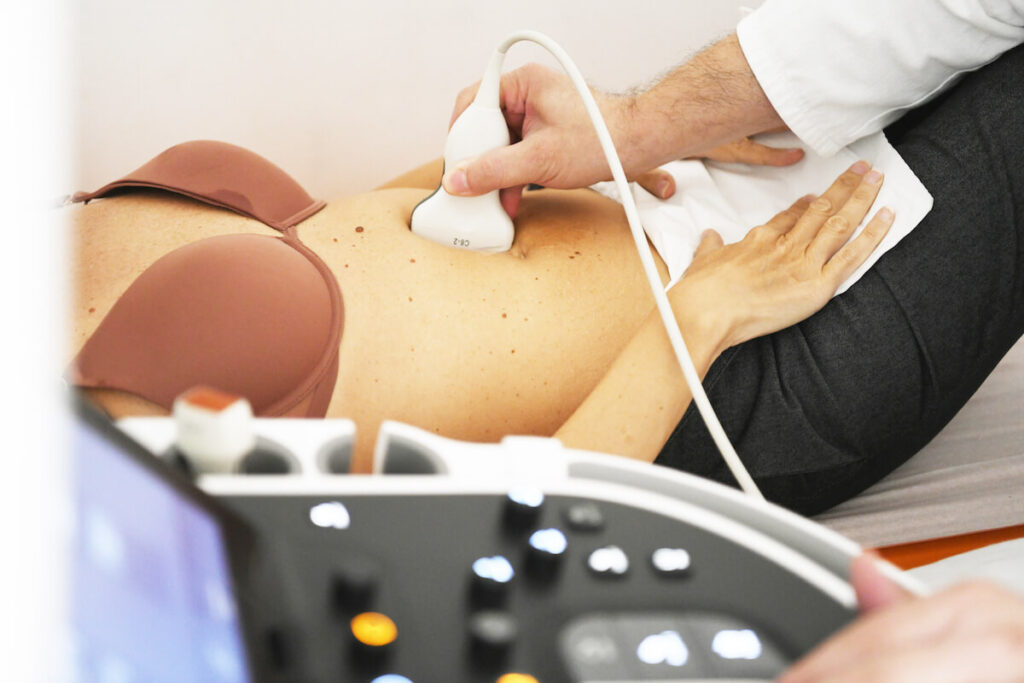
What to expect during abdominal aorta doppler?
Doppler ultrasound of the abdominal aorta is usually a painless and non-invasive procedure that takes a relatively short time, and depends on the specific needs and goals of the examination, as well as on the speed and experience of the ultrasound technician performing the examination. Typically, this examination takes between 15 and 45 minutes.
During the Doppler ultrasound of the abdominal aorta, you can expect the following steps and situations:
- Preparation: Before the actual examination, the medical technician or ultrasound technician will invite you to lie down on the examination table. They will provide you with a comfortable position and cover you with a sterile drape.
- Ultrasound gel: The technician will apply ultrasound gel to the skin where the ultrasound probe (transducer) will be placed. The gel enables better conduction of sound waves between the skin and the probe.
- Probe examination: The ultrasound probe will be placed on the skin at a specific point on the abdomen or back, depending on the purpose of the examination. The probe is connected to a computer that receives and processes information.
- Ultrasound waves: The technician will use a probe to emit ultrasound waves through the skin into the body. These waves are reflected from internal organs and blood vessels, and their echoes are registered and converted into images and sound signals on the screen.
- Moving the probe: The technician will move the probe over the examined area to get different views and information. You may be asked to change your body position to allow better access to certain parts of the abdominal aorta.
- Blood flow monitoring: Doppler technique will be used to monitor the speed and direction of blood flow through the abdominal aorta and its branches. This will allow you to assess whether there is any abnormality in blood flow, such as narrowings or aneurysms.
- Sound and images: During the examination, you may hear sounds that represent blood flow, and you will see images on the screen that show the structure and blood flow of the abdominal aorta.
- Completion of the examination: When the technician has finished collecting all the necessary information and images, he will remove the gel from the skin and inform you that the examination is complete.
- Continuation of treatment: After completing the examination, you can get dressed and continue with your daily activities. The technician will forward the obtained information to the doctor for analysis and diagnosis.
- After the results are obtained, the doctor will analyse the information obtained and discuss with you the results and possible further steps in the treatment or monitoring of your medical condition.
Abdominal aorta doppler – Price:
The price of this procedure is 7.900 RSD.