What is MSCT of the abdomen and pelvis
CT examination or scanning of the abdomen and pelvis is a painless radiological method of examination, which is used to make a series of cross-sectional images of this part of the body (where bones, blood vessels and soft tissue can be clearly seen), and uses computer technology and X-rays to achieve accurate and clear shots.
MSCT images of the abdomen provide detailed information about all the organs that are located in the abdominal cavity and provide a detailed overview of any potential injuries or diseases that affect these organs. It is also used to detect the causes of unexplained abdominal pain or in emergencies, with the help of these images, internal bleeding and organ injuries can be detected.
A CT scanner provides much more information and more accurate images than X-rays, which is why it is used to monitor the size and condition of tumors, then to scan the pancreas, liver, gallbladder, kidneys in many other cases. When it comes to the pelvic part, CT scanner can take images of female and male reproductive organs (uterus and ovaries, or prostate and seminal vesicles in men), but also the bladder, the final parts of the ureter, as well as parts of the musculoskeletal system, vascular structures and fig.
MSCT abdominal scanning can also be used to visualize needle placement during abdominal or tumor biopsies or during aspiration (withdrawal) of fluid from the abdomen. MSCT images of the abdomen are useful in monitoring tumors and other conditions of the abdomen before and after treatment.
Other related procedures that can be used to diagnose abdominal problems include abdominal X-ray, pancreatic scanner, liver scanner, gallbladder scanner, kidney scanner, endoscopic procedures such as colonoscopy, abdominal ultrasound, and abdominal angiogram.
Multi-slice computed tomography – MSCT of the abdomen and small pelvis, with intravenous CT urography is a diagnostic method of contrast imaging of the kidneys and urinary tract (ureter, bladder and urethra), which allows insight into kidney function and anatomical structure of the urinary tract.
Indications for MSCT of the abdomen and pelvis
This procedure is used when a patient reports to a doctor with pain in the abdomen or pelvis, which cannot be diagnosed in any other way and the cause of the pain cant be discovered. Also, when the presence of free fluid is determined, or when an injury or fracture of certain bones in this part of the body is suspected.
CT is very useful when it comes to imaging the abdomen and pelvis, if doctor is looking of some kind of benign or malignant tumor (oncological urogenital and gastroenterological pathology), as well as inflammatory and surgical urogenital pathologies, such as inflammation of the bladder and urinary tract, postoperative complications such as the appearance of blood in the urine and its swelling outside the urinary tract. Scanning is also recommended for gastroenterological patients, who have problems with inflammatory bowel diseases or tumor changes.
This method can also detect the appearance on the pancreas and its inflammation, also known as pancreatitis, then it is used to diagnose cirrhosis of the liver, liver cancer, kidney cancer, ovary cancer , kidney stones or bladder. In addition, injury to all internal organs (from the liver, through the spleen, all the way to the kidneys) can be easily detected using an MSCT scanner.
How to prepare before CT scan of the abdomen?
The patient is referred to the CT scan by a specialist, one of the following branches of medicine: urology, gynecology, abdominal or vascular surgery and oncology. In order to make the scan as accurate as possible, it is necessary to submit medical documentation (creatinine and urea test not older than 15 days, due to possible contrast; reports of specialist which will show why the patient was referred for CT scan), which will provide the necessary information to the radiologist to perform as accurate scan as possible.
If the patient uses therapy to regulate blood sugar, it is necessary to stop taking the drug the day before the recording and on the day of the recording. In the case if patient is allergic to iodine or has allergic reactions to certain foods such as eggs, nuts and fish, he should inform our medical staff, so that an allergist can be involved in further preparations. He could give the patient certain medications, in order to prepare him for possible contrast and prevent him from getting an allergic reaction.
Females are warned that if they are pregnant or suspect they may be, they should inform their doctor, because the X-ray can negatively affect the fetus, so it is necessary to determine some other type of diagnosis such as magnetic resonance imaging or ultrasound examination.
If you have any additional questions or concerns, you can certainly ask our doctors during the process of preparation for the recording and they will give you all the information important to you, but also to them, so that the examination went as well as possible and the results were as accurate as possible.
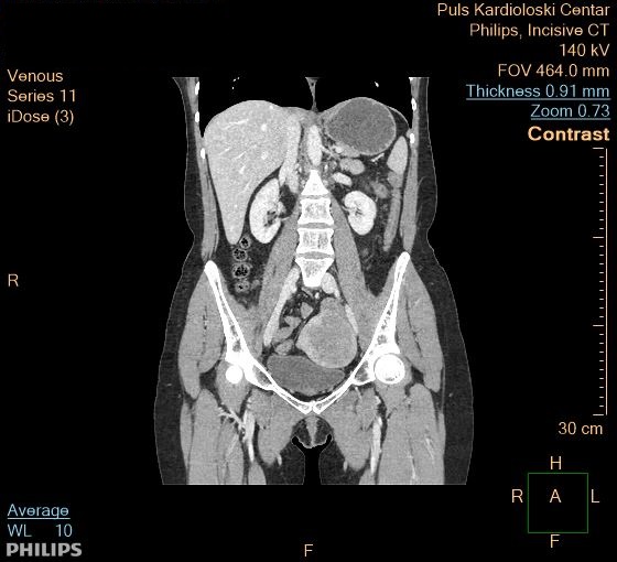
CT scan at the Pulse Cardiology Center
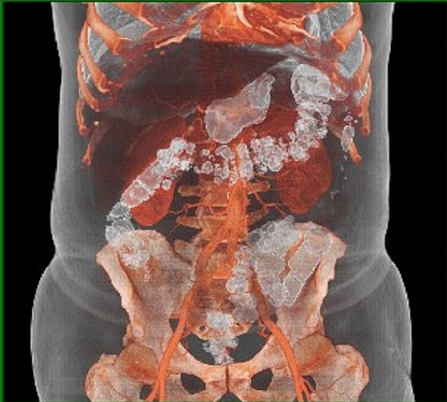
During the examination, the patient lies on a narrow platform in the shape of a table, which occupies the central part of the CT scanner and is usually placed in a lying position with his arms raised above his head. The scanner is open on both sides, so the patient can communicate with the radiology technician and other staff. When the scan starts, the CT scanner starts to scan the abdominal part of the body, by making a larger number of images as it rotates around the patient. By merging all these images, a three-dimensional representation of the internal organs was obtained, which can be viewed on the screen, but also printed if needed.
Sometimes it is necessary for the patient to receive contrast intravenously, in order for certain organs to be better seen on the image. Contrast is a compound that helps the organs to be seen more clearly in the image, by coloring them white.
If the patient has ever had a reaction to a contrast agent or has a condition that may be contraindicated for imaging in this way, he should tell the doctor, so that all measures can be taken in time. At the moment of receiving the contrast, the patient can feel a slight tingling, metallic taste in the mouth, a rush of heat in the body and all these symptoms are expected and do not last longer than a few moments.
The recording lasts between 15 and 30 minutes, after which the patient is advised to drink as much fluid as possible during the day, if he has received contrast, in order to speed up his excretion through the kidneys. After the recording, the patient returns to normal activities.
What are the risks of MSCT scanning
You may want to ask your doctor about the amount of radiation used during the MSCT procedure and the risks associated with your particular situation. It is a good idea to keep a record of past radiation exposure, such as previous MSCT images and other types of X-rays, so you can tell your doctor. The risks associated with radiation exposure may be related to the cumulative number of X-ray examinations or treatments over a long period of time.
If you are pregnant or suspect you may be pregnant, tell your doctor. Radiation exposure during pregnancy can lead to congenital damage to the fetus.
If a contrast material is used, there is a risk of an allergic reaction. Patients who are allergic to or sensitive to drugs should inform their doctor. You will need to tell your doctor if you have ever had a reaction to contrast material or kidney problems. If you are taking drugs from the biguanide group or similar medicines, you may be asked to stop taking the medicine at least 48 hours after applying the contrast, as this may cause a condition called metabolic acidosis or an unsafe change in blood pH.
Patients with kidney problems should inform their doctor about the diseases. In some cases, contrast material can cause kidney failure, especially if the person is dehydrated or already has underlying kidney disease.
There may be other risks depending on your specific health condition. Be sure to talk to your doctor before the procedure.
Why should you schedule an examination at the Pulse Cardiology Center?
Many years of experience and highly trained staff are our best recommendation. Our experts work on the most modern devices, so it is not surprising that they achieve top results in the treatment of patients. A professional team of radiologists is ready to answer all patients’ questions and provide a detailed and precise interpretation of the CT scanner results. After that, radiologists will also recommend in which direction the treatment should proceed.
Patient care is at the top of our list of priorities, so make an appointment at the Pulse Cardiology Center in New Belgrade and make sure of the top quality of our services. We try to understand your health problems and we pay the highest level of attention to each patient from the first moment, in the desire to find a solution as soon as possible and establish a timely diagnosis.