Home » Services » Ultrasound (Ultrasound Diagnostics) » Doppler of Legs Blood Vessels
Doppler of Legs Blood Vessels
What is vascular doppler of the legs?
Doppler of the leg blood vessels (also known as Doppler ultrasound) is a medical procedure used to evaluate blood circulation in the blood vessels of the legs. This procedure uses the Doppler effect, which is based on the change in frequency of sound waves when they are reflected by moving objects, such as red blood cells in the blood.
Color Doppler of leg blood vessels is an advanced technique that is used together with standard Doppler ultrasound to further improve the diagnostic value of the examination. This method combines traditional Doppler ultrasound with color display of blood flow, which allows color visualization and analysis of blood circulation on the screen.
Leg Doppler is often used to diagnose blood circulation problems, such as narrowing of the arteries (atherosclerosis), blood clots, thrombosis, varicose veins, or other vascular abnormalities. It can also be used in people who have leg pain, swelling or other symptoms that indicate vascular problems.
This procedure is non-invasive, painless and safe, so it is often used as the first step in diagnosing diseases of the blood vessels of the legs. Based on the results of the Doppler ultrasound, the doctor can recommend further tests or therapy in order to adequately treat the problem of blood circulation in the legs.
Why is color doppler blood vessels of the legs used?
Here are some reasons why color doppler of the blood vessels of the legs is used:
- Better visualization of blood flow: Color Doppler allows doctors to see blood flow in arteries and veins in color, making it easier to identify changes in flow, narrowings, blockages, blood clots or other vascular abnormalities.
- More precise analysis: Color Doppler allows doctors to more precisely determine the speed and direction of blood flow in different blood vessels of the legs, which is especially useful when monitoring changes over time or after interventions.
- Faster diagnosis: The use of color doppler can speed up the diagnostic process, as it allows doctors to quickly identify problems in blood circulation in the legs, which can be of critical importance in emergency situations such as thrombosis or acute occlusion of arteries.
- Improved communication with patients: Color Doppler allows doctors to show patients more clearly the state of their blood vessels, which can improve patients’ understanding of their health condition and proposed treatment.
- Monitoring the progress of therapy: Color Doppler is often used to monitor the effectiveness of therapy in leg vascular disease, to determine whether improvement has occurred.
- In peripheral artery disease, also known as peripheral arterial disease (PAB), which is characterized by narrowing or blockage of the arteries that supply blood to the extremities, such as the legs, color Doppler facilitates the identification of problems with blood flow in the arteries: artery stenosis (narrowing), artery occlusion, development of collateral circulation, tissue perfusion
- Color Doppler of leg blood vessels after stent or graft implantation is a diagnostic procedure used to monitor blood flow and functioning of implanted stents or grafts in leg arteries. Color Doppler facilitates the identification of possible problems with the implanted stent or graft, such as: stenosis or restenosis, thrombosis, movement or leakage of the stent or graft from its position and intimal hyperplasia, which is an abnormal growth of the inner layer of the artery around the stent or graft. layer of the artery around the stent or graft.
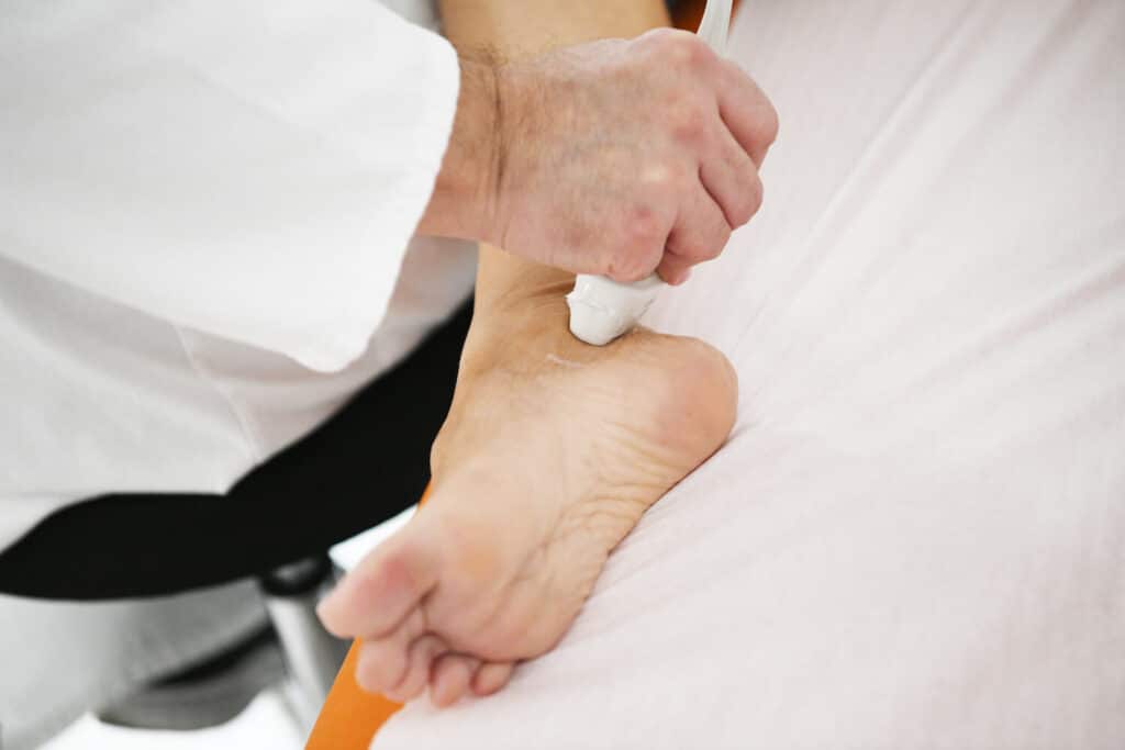
What does color doppler of the blood vessels of the legs mean? What happens during the review?
Color Doppler of leg blood vessels is a non-invasive medical procedure that involves the following steps:
- The patient will be asked to prepare beforehand for the examination. This may involve removing clothing from the area to be examined (usually the legs) and putting on a medical gown.
- The patient will be placed on a suitable table or bed so that the legs are accessible to the ultrasound probe. It is necessary to ensure that the patient is comfortable during the examination.
- The doctor or technician will apply the gel to the skin of the legs in the area to be examined. The gel helps to improve the contact between the skin and the ultrasound probe to better transmit the sound waves.
- The doctor or technician will use an ultrasound probe (in this case color Doppler) and carefully move it over the surface of the skin of the legs. The probe emits sound waves and captures their reflections from blood vessels.
- An image of the blood vessels will be displayed on the screen, and the colors will indicate the direction and speed of the blood flow. Blue color is usually used for blood flow towards the heart (in veins) and red color for blood flow away from the heart (in arteries).
- The doctor will carefully analyze the blood flow image to assess the speed, direction and smoothness of the blood flow in the arteries and veins of the legs. Based on this information, the doctor can detect possible problems, such as narrowings, blockages, blood clots or varicose veins.
- Based on the results of the color doppler examination, the doctor will make a diagnosis and, if necessary, recommend further diagnostics or treatments for the patient.
- After the examination, the gel will be removed from the skin, and the patient can continue with his daily activities normally.
It is important to note that color doppler of the blood vessels of the legs is a safe and painless procedure, and the results of the examination can be of great help in the diagnosis and monitoring of various vascular conditions and diseases.
How should you prepare for color doppler of leg blood vessels?
Preparation for color Doppler blood vessels of the legs is usually simple and does not require special dietary measures or procedures. However, it is important to follow the instructions of the healthcare staff to ensure the best results from the examination. Here are some common preparation guidelines:
- Inform the health care professional about medications: Before starting the examination, tell the health care professional about all the medications you are taking. Some medications, such as blood thinners, can affect blood circulation and test results.
- Comfortable clothing: Make sure you wear comfortable and loose clothing that will be easy to remove from the area to be examined (usually the legs). The medical staff will tell you if you need to wear a special medical gown or sleeves for the examination.
- Removal of jewelry: Before the examination, you should remove jewelry, such as bracelets, rings, or necklaces, from the area to be examined. Jewelry can interfere with the ultrasound and make it difficult to get clear images.
- Pay attention to eating habits: In most cases, there are no special restrictions regarding food and drink before the color Doppler examination of the blood vessels of the legs. However, if there are special requirements, the health staff will tell you.
- Medical information: If you have a previous diagnosis or history of circulation problems, thrombosis or other vascular conditions, be sure to inform the health care staff. This will help them tailor the review to your individual needs.
- Follow the instructions: If the health care staff gives you special instructions or preparation guidelines, be sure to follow them to ensure the best results of the examination.
It is important that you do not hesitate to ask questions or express your concerns to the healthcare staff before the examination. They are here to help you and provide you with the best possible care during the Color Doppler leg blood vessel procedure.
How long does the examination take?
The duration of a color Doppler examination of the blood vessels of the legs can vary depending on several factors, including the complexity of the examination, the information required and the individual characteristics of the patient. Typically, this examination takes between 30 and 60 minutes. There are several factors that can affect the duration of the examination:
- If only a specific part of the leg needs to be examined, the examination will usually take less time. However, if the entire leg or multiple areas need to be examined, the examination may take longer.
- If there are suspicions of more serious vascular problems or there is a need for a more detailed analysis, the examination can be extended to comprehensively assess blood circulation.
- If the patient is calm and cooperative during the examination, it can speed up the process and shorten the examination time. However, if the patient is restless or requires additional time to set up and get comfortable, the examination may take longer.
- After the examination is completed, the doctor will analyze the collected data and make a diagnosis. The time required to interpret the results may vary depending on the complexity of the findings.
It is important for patients to be patient during the examination, as precision and detail are key to making an accurate diagnosis.
What problems or conditions can color doppler of the blood vessels of the legs show?
Doppler blood vessels of the legs can detect various problems or conditions in the blood circulation of the lower extremities. Here are some common problems or conditions that can be identified with a color Doppler examination of the blood vessels of the legs:
Atherosclerosis (clogged arteries)
This is a condition in which the arteries gradually narrow due to the accumulation of fatty deposits and plaque on the inner walls of blood vessels, which can reduce blood flow and lead to peripheral artery disease (PAD).
Peripheral Arterial Disease (PAD)
PAD is a condition in which the arteries in the legs are narrowed or blocked, which can lead to symptoms such as pain when walking (claudicatio intermittens) or reduced blood flow to the legs.
Blood clots
Thrombosis is the formation of a blood clot inside a blood vessel, while embolism involves the release of a clot that can be carried by the bloodstream to other parts of the body. Both conditions can be serious and require immediate attention.
Venous insufficiency
This abnormality refers to the improper functioning of the veins, which can lead to problems with blood returning from the legs to the heart. Color Doppler can detect enlarged veins (varicose veins) and assess blood flow in the veins to determine insufficiency.
Complications of arteriovenous fistulas or grafts
Doppler of the blood vessels of the legs is also used to monitor the function of arteriovenous fistulas or grafts, which are artificial pathways used for hemodialysis in patients with kidney disease.
Vasculitis
It is an inflammatory condition of the blood vessels that can affect the blood flow in the legs.
Aneurysms
An aneurysm is an enlargement of an artery, which can be dangerous because there is a risk of rupture. Color Doppler can detect aneurysms in the arteries of the legs.
Complications after vascular interventions
Color Doppler can be used to evaluate the condition after vascular interventions, such as angioplasty, after stenting, grafting or other vascular operations.
These are just a few of the many conditions and problems that a color Doppler examination of the blood vessels of the legs can identify. Based on the results of this examination, the doctor can make an accurate diagnosis and recommend the appropriate treatment in order to adequately manage the problem in the blood circulation in the legs.
Who should examine the test results?
The results of the color doppler examination of the blood vessels of the legs are examined by a medical specialist, usually a vascular surgeon, a vascular radiologist, or an ultrasound specialist (a radiologist with experience in ultrasound diagnostics). These specialists have training and experience in interpreting ultrasound images and Doppler blood flow analysis.
After the color Doppler examination of the blood vessels of the legs is completed, the medical technician who performed the examination prepares the images and findings, and then forwards them to the specialist for interpretation. The specialist will carefully analyze the collected data and prepare a report with a conclusion, in which he will state his diagnostic findings and recommendations for further action.
Next, the patient will have an appointment with a specialist who will convey the results of the examination, explain the diagnosis and discuss possible treatment options, if necessary. The specialist will also answer questions and provide additional information about the patient’s health.
It is important that the patient has confidence in the expertise and experience of the specialist who interprets the results of the color Doppler examination of the blood vessels of the legs, as this can be crucial for an accurate diagnosis and proper treatment.
What additional tests can the doctor recommend based on the color doppler results of the blood vessels of the legs?
After a color doppler examination of the blood vessels of the legs, if there are suspicions of certain problems or the need for additional evaluation, the doctor may recommend additional diagnostic tests. This can vary depending on specific symptoms and examination findings:
Ankle-brachial index (ABI) test:
This is a simple test used to assess the severity of peripheral arterial disease (PAD). The pressure on the arm and ankle joint is measured, and based on the ratio of these values, the degree of narrowing of the arteries in the legs can be estimated.
Duplex ultrasound combines color Doppler with B-mode ultrasound to provide more detailed information about the structure of blood vessels and the presence of plaques or other changes in the vessel walls.
Angiography
Angiography is an invasive procedure that uses X-rays to visualize blood vessels. It can be used for a more detailed assessment of the condition of the arteries and veins of the legs, especially in complicated cases.
CT angiography (CTA) or MR angiography (MRA)
These noninvasive imaging techniques use CT or MRI technology to produce three-dimensional images of blood vessels and blood flow in the legs.
Coagulation tests and hematological tests
If thrombosis is suspected, coagulation tests (eg, D-dimer test) and hematological tests may be ordered to assess the risk of thrombosis.
Functional tests
In some cases, a doctor may recommend functional tests that assess muscle and circulatory performance during certain activities, such as a walking test.
It is important for the patient to discuss any suspicions, symptoms or questions they have with their doctor so that the doctor can recommend appropriate additional tests that will contribute to an accurate diagnosis and appropriate treatment planning.
Doppler of legs blood vessels – Price
Price for this kind of exam is 7.900 RSD.