What is an X-ray?
X-ray imaging, or radiography, is a diagnostic procedure that uses X-rays to create images of the internal structures of the body. This non-invasive method is widely used in medical diagnostics to obtain information about tissues and organs.
Advantages of X-rays
Here are several key advantages of X-ray diagnostics:
- Speed: X-ray images provide rapid diagnosis because images can be obtained in real time. This quick availability of information assists doctors in emergency situations and in making immediate medical decisions.
- Wide applicability: X-rays can be used to examine various parts of the body, including bones, lungs, joints, abdominal organs, and many other structures.
- Low cost: X-ray is a cheaper method compared to magnetic resonance imaging (MRI) and CT scanners, and it is widely available.
- Monitoring treatment results: X-ray images are often used to monitor the progress of treatment, especially in patients with injuries or bone diseases.
- Minimal invasiveness: X-ray imaging is a non-invasive, painless method.
Significance of X-rays
Advancements in X-ray technology not only improve diagnostic capabilities but also enhance patient comfort, reduce radiation exposure, and facilitate the work of medical staff. Imaging software increasingly contributes to improving the quality of X-ray images, enabling clearer visualization of organs, tissues, and bone structures, as well as easier identification of pathologies if present. Therefore, X-rays are still considered a highly useful diagnostic method, with the greatest significance and application found in the following areas:
- Diagnosis and disease monitoring: X-ray imaging enables visualization of internal structures, which is crucial for diagnosing various medical conditions, including fractures, tumors, infections, and lung diseases.
- Surgical planning: Surgeons often use X-ray images to plan surgeries, allowing for a more precise approach and reduced risks during procedures.
- Treatment monitoring: X-ray images allow monitoring the progress of diseases or injuries over time, which is important for adjusting therapy and assessing treatment success.
- Early disease detection: X-ray imaging can help detect diseases in their early stages, enabling faster and more effective treatment.

Body parts most commonly imaged with X-rays
X-ray of the head (skull radiography)
Often performed after head injuries such as blows, falls, or traffic accidents to rule out fractures or other injuries. If there is suspicion of a skull bone fracture, X-ray imaging can help confirm the diagnosis and determine the severity of the injury. Head imaging is often done in cases of headaches, dizziness, loss of consciousness, or neurological symptoms. It is also used to monitor bone development in children, especially in cases of suspected bone pathologies or cranial anomalies.
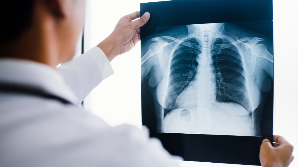
Lung X-ray
Also known as chest radiography, is often prescribed in cases of respiratory complaints such as cough, difficulty breathing, chest pain, or other breathing problems, pneumonia (lung inflammation), bronchitis, lung tumors, and assessment of potential changes in lung circulation. It is also used to control and monitor asthma, chronic obstructive pulmonary disease (COPD), and interstitial lung diseases. Before certain types of surgeries or medical procedures, chest X-ray imaging may be part of the preoperative assessment to rule out potential complications or abnormalities.
Chest X-ray
It is a diagnostic procedure used to visualize structures in the chest, including the lungs, heart, bones, and diaphragm. It is used for diagnosing chest trauma and fractures, lung diseases, and also serves as part of the evaluation of cardiac issues, especially for determining heart size
Abdominal X-ray native imaging
It is used to visualize internal structures in the abdomen, including organs such as the stomach, intestines, liver, spleen, and kidneys. This examination is applied in certain clinical situations for diagnosis or monitoring of certain conditions such as: abdominal pain, intestinal obstruction, kidney stones, or other structural abnormalities. It is used to detect and monitor gastrointestinal problems, including bloating, presence of free gas, or foreign bodies in the intestines. When there is suspicion of constipation or ileus (lack of normal bowel movements), native abdominal imaging can help identify the causes and determine the degree of obstruction. It is used to monitor chronic bowel diseases such as Crohn’s disease and ulcerative colitis. Native abdominal imaging can be applied after trauma or injury to the abdominal cavity to rule out possible bone fractures, internal organ injuries, or presence of free fluid. After abdominal surgeries, it can be used to monitor postoperative changes and identify any complications.
Urinary tract X-ray – native imaging
It is used to visualize the kidneys, ureters, urinary bladder, and urethra. It is used in the presence of the following symptoms: abdominal or lower back pain, frequent urination or painful urination, blood in the urine. These symptoms may indicate urinary tract infection, presence of stones, tumors, or other disorders. Native urinary tract imaging may be recommended for monitoring congenital anomalies. Before surgical interventions in the urinary tract area, the doctor may recommend native imaging.
X-ray of the paranasal sinuses
It may be recommended to diagnose sinusitis (sinus inflammation), tumors, cysts, or other abnormalities in the paranasal sinuses and identify the causes of facial or head pain. X-ray imaging can help assess the anatomical structure of the paranasal sinuses, which is useful in planning surgical procedures or other medical interventions as well as in identifying potential causes of recurrent sinus infections, such as anatomical variations or other structural changes.
Nasal X-ray (AP + LL) nasal bone
It including anteroposterior (AP) and lateral (LL) nasal bone imaging, can help assess possible fractures of the nasal bones or other injuries, for the analysis of anatomical changes and deformities of the nose including septal deviations or other structural abnormalities, diagnosis of the causes of breathing difficulties, assessment of nasal passage patency, which is useful in cases where there is suspicion of obstruction, narrowing or infection of the nose. Before some nasal surgeries, the doctor may recommend X-ray imaging for a better understanding of the anatomical structure and planning of the surgical procedure.
Spinal X-ray
It is performed to assess the condition of the spinal column. If there is suspicion of spinal injury due to accidents, falls, or other traumatic events, X-ray imaging can help in the diagnosis of fractures, dislocations, or other serious injuries. X-ray is often used to assess spinal deformities, such as scoliosis (curvature of the spine sideways), lordosis (increased curvature forward), or kyphosis (increased curvature backward). X-ray imaging can be used to assess degenerative changes in the spine, including arthritis, spondylosis, or other degenerative spine diseases. Neurological symptoms such as tingling, weakness, loss of sensation, or other manifestations indicating possible compression of nerve structures in the spine are also indications for spinal X-ray.
X-ray of the cervical spine
It is usually performed to detect the causes of neck pain or neurological symptoms (tingling, weakness in the arms), trauma, arthritis, and degenerative changes in the cervical spine.
X-ray of the thoracic spine
It is performed to detect the causes of back pain, deformities, and spinal trauma. X-ray imaging can be used to identify suspicious lesions or tumors in the thoracic spine, which is especially important in the diagnosis and monitoring of malignant processes.
X-ray of the lumbar spine
It may be recommended to identify the causes of pain, including fractures, degenerative changes, herniated discs, or other pathologies.
Bone and joint X-ray
It is the method of choice for identifying bone fractures after injuries. X-ray imaging can be used to assess the anatomy of bones and joints to identify any deformities, including scoliosis, flat feet, or other structural changes. In cases of suspected arthritis, X-ray examination can help detect characteristic changes in the joints, such as osteophytes (bone spurs), narrowing of the joint space, or bone erosions. It is used for the diagnosis of bursitis (inflammation of bursae (fluid-filled sacs) and tendonitis (inflammation of tendons).
Oncological imaging of 11 images
It is performed to detect metastatic changes in bones, upon request of the competent specialist.
How to prepare for an X-ray examination?
Preparation for an X-ray examination varies depending on the type of examination and the part of the body to be imaged. Follow the instructions of our medical staff regarding any special requirements or preparations for your specific examination.
- If you are pregnant or suspect you are pregnant, be sure to inform the medical staff before the X-ray examination. X-ray radiation can affect the fetus, so it is important to consider this information when planning the examination.
- Before the examination, remove jewelry, watches, clasps, and other metal objects that could interfere with imaging or cause artifacts on X-ray images. Wear clothing that does not contain metal parts to avoid interfering with X-ray radiation. If you do not follow these recommendations, the image will not be accurate and the doctor will not obtain all the necessary information.
- If you have allergies, especially to contrast agents that are sometimes used, inform the medical staff in advance. Certain types of X-ray examinations may require special preparation. For example, for X-ray imaging of the digestive tract (stomach), contrast material may be required.
What does the imaging look like?
The technician will try to position the patient so that the best possible images are obtained. Then, they will protect the patient with a lead vest so that parts of the body not being imaged are not exposed to unnecessary radiation. The patient will be instructed to rest and maintain a certain position during the imaging procedure. To achieve the best results, the patient may need to hold their breath for a few seconds. This helps reduce the possibility of movement that could blur the image. In some cases, it may be necessary to image a body part from different angles to obtain more precise diagnostic information.
To ensure your safety, our staff maintains a special relationship with patients:
- Patient communication: During the procedure, communication with the patient is crucial. The staff will explain each phase of the procedure, provide guidance, and ensure that the patient is informed and feels safe.
- Application of anxiety reduction techniques: X-ray procedures can cause anxiety in some patients. Our staff is trained in anxiety reduction techniques such as talking to the patient during the procedure or providing additional time for preparation.
- Equipment for ensuring stability: If necessary, patients can be provided with appropriate pillows, supports, or other types of equipment to maintain stability and comfort during X-ray imaging.
- Meeting special needs: The staff takes into account the special needs of patients, including pregnant women, people with disabilities, or those with special medical requirements
Interpretation of results
Radiologists analyze the images to identify anatomy, assess structure, and confirm or rule out suspicion of any changes or pathologies. Based on the analysis of X-ray images, the radiologist prepares a report containing a description of the findings and diagnoses. The doctor who referred the patient for X-ray examination reviews the report and uses it for further patient treatment.
The timeframe for obtaining X-ray examination results may vary: in emergency cases, results may be available very quickly to enable prompt medical intervention. In routine X-ray examinations, the result will arrive on the same day.
Limitations of X-ray imaging
- No information about soft tissues: X-ray imaging is particularly good for visualizing bones, but it does not provide detailed information about soft tissues such as muscles, organs, or blood vessels. The method of choice for these structures is magnetic resonance imaging (MRI) or CT scanners.
- Not used during pregnancy: Although modern X-ray procedures are usually applied with minimal radiation dose, their use during pregnancy is avoided to reduce fetal exposure to X-ray radiation.
- Not suitable for detecting soft tumors: X-ray images are not ideal for detecting small or soft tumors, especially in early stages. Scanners or magnetic resonance imaging are often more effective for these purposes.
- Does not provide three-dimensional representation: X-ray images offer a two-dimensional representation of structures, which can make precise assessment of some anatomical characteristics difficult.