What Is a Carotid-Cavernous Fistula?
Carotid cavernous fistula is a condition that affects your eye. It can come on suddenly after a head injury or slowly over time. When an abnormal connection happens between either of the carotid arteries and the veins just behind the eye, it is called a carotid cavernous fistula.
Because the carotid arteries have a greater pressure of blood flow than do the veins, the rush of blood prevents the veins that serve the eyes from draining properly.
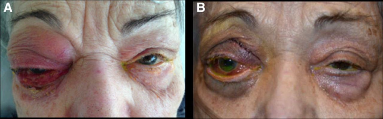
The blood pressure starts to build up in the eye, causing it to bulge. The eye turns red. As the condition gets worse, a person may start losing vision. Without treatment, the sight can be lost in that eye.
What Causes a Carotid-Cavernous Fistula?
A carotid-cavernous fistula is an irregular connection between your carotid artery and a vein called the cavernous sinus. The cavernous sinus is behind your eyes and drains blood from your facial veins.
There are two main types of carotid-cavernous fistulas:
Direct. This type is sometimes also called trauma carotid cavernous fistula. It is a hole or tear in a branch of your carotid artery inside the cavernous sinus. This tear is called a fistula, and it is usually caused by accident or injury, including:
- Being hit in the head
- Car accident
- Fall
- Penetrating head injury like a stabbing
- It can also be iatrogenic, which means it happens as a complication from surgery or treatment.
Indirect. This occurs when there is an irregular connection between the carotid artery and the cavernous sinus.
It’s not exactly clear why it happens, but sometimes the artery, or a branch of the artery, will reroute and the blood flow changes. This rerouting is called a shunt.
A spontaneous carotid fistula can happen because of a blockage in the artery or other conditions, including:
- Ruptured aneurysm
- Ehlers-Danlos syndrome
- Pregnancy
- Straining
- Atherosclerosis, or hardening of your arteries
When a fistula happens, high blood flow from the artery is pushed into a low pressure cavernous vein. This causes problems with blood drainage from your eye socket and can cause your eye to bulge.
It can sometimes be confused for thyroid eye disease where an overactive thyroid causes your eyes to bulge.
Acquired rather than congenital vascular malformations, carotid-cavernous fistulas (CCFs) may arise spontaneously or from secondary causes. CCFs can present with a variety of signs and symptoms. Many lesions are associated with significant neuro-ophthalmologic morbidity and mortality. Treatment decisions require multiple considerations: the nature of the symptoms, the location of the lesion, the complexity of the angioarchitecture, and the risk of visual and neurologic morbidity.
Carotid Cavernous Fistula Symptoms
Direct carotid fistulas usually happen within a few days or weeks of having a head injury. Symptoms can include:
- Red eyes
- Bulging eye
- Blood flow sounds around your eye
- Pulsing eyeball
- Eye pain
- Weak or paralyzed eye muscle
- Drooping eyelid
Indirect carotid fistulas usually come on slowly over time. Symptoms can include:
- Mild eye bulging
- Eye congestion
- Headache
- Pulsing eyeball
Sometimes it’s also hard to close your eyes, which can cause dryness. If your eyelid is drooping, it might be hard to see, too.
Diagnosis of a Carotid Cavernous Fistula
Your doctor will take your personal history and do a physical examination. They will also order some imaging tests to see what’s happening inside your head and eye. These can include:
Cerebral angiogram. A cerebral angiogram is an X-ray of your head and neck. It looks for any blockages or other irregularities in your blood vessels.
Pneumotonometry. This test measures the pressure inside your eyes by exposing it to a sudden puff of air.
Doppler ultrasonography. Your doctor will use a Doppler ultrasound to listen to the blood flow in your eye socket.
Magnetic resonance imagery. Also called an MRI, this imaging test will help your doctor see your veins, arteries, and your brain.
Cerebral angiography is the gold standard for the definitive diagnosis, classification and treatment planning of these lesions.
Treating a carotid cavernous fistula with embolization involves placing small platinum coils where the abnormal connection is. This separates the blood flow of the carotid arteries from that of the veins. As a result, the blood can drain properly from the eyes.
Endovascular treatment of Carotid-Cavernous Fistula
Endovascular treatment of Carotid-Cavernous Fistula in a minimally invasive way through the inguinal artery leads to the fistula itself, which gives the interventional neuroradiologist a much clearer picture of the fistula itself, size, location, venous drainage and the presence of main venous collaterals. After that, the interventional neuroradiologist decides which method to perform the embolization and close the fistula.
Coil embolization is done in an angiography suite. Like other endovascular procedures, this one is performed under local anesthesia, and complications are reduced to a minimum and the patient is discharged from the hospital after 24-48 hours. The procedure doesn’t require any incision into the head or skull.
During the procedure, a small tube is placed through an artery in the groin. The tube is advanced up to the arteries in the neck. Another smaller tube is threaded through the first one. Small platinum coils are delivered to the abnormal connection through the second tube. The coils separate the blood flowing in the arteries from the blood flowing in the veins.
Coil embolization treatment often completely reverses the disabling symptoms in the eye, if treated early enough.
Risks of a Carotid-Cavernous Fistula
The prognosis for a carotid cavernous fistula is generally good. It isn’t considered life-threatening, but you should have your eye treated right away to protect your eyesight and eye health.
It can take a few weeks to months for the fistula to fully close and for blood flow to change. Once it is closed, symptoms improve within hours to days.
Most people with indirect fistulas are better within 6 months of treatment. Sometimes people who have a direct fistula have lasting side effects, including:
- Bulging eye
- Vision loss
- Muscle weakness in the eye
- Muscle paralysis in the eye
If you’re experiencing changes in your eye or eyesight, talk to your doctor.
A Rare but Treatable Cause of Rapidly Progressive Vision Loss
Carotid cavernous fistulas (CCFs) are a rare but potentially devastating cause of orbital symptoms, visual loss, and periocular disfigurement.
Carotid cavernous fistula patients typically present with proptosis, elevated intraocular pressure, prominent tortuous conjunctival vessels, and sometimes headache.
Endovascular treatment is the modality of choice for carotid cavernous fistulas. Prompt treatment can prevent further complications of carotid cavernous fistulas and lead to complete resolution of symptoms.
Case report:
After the traffic accident, the patient complains of noise in the head, pulsation of the right eyeball, redness of the eye and prominence of the eyeball. Le DSA was performed, where carotid-cavernous fistula was proven. The fistula is closed by the endovascular treatment.
After the intervention, the eyeball completely came back, and the blood supply to the sclera receded.