Why is CT scanner the method of choice for diagnosing coronary artery disease?
Until recently, magnetic resonance imaging (MRI) was the preferred method for imaging stable coronary artery disease. However, CT scanning has proven to be a more advanced way to assess the patient’s condition.
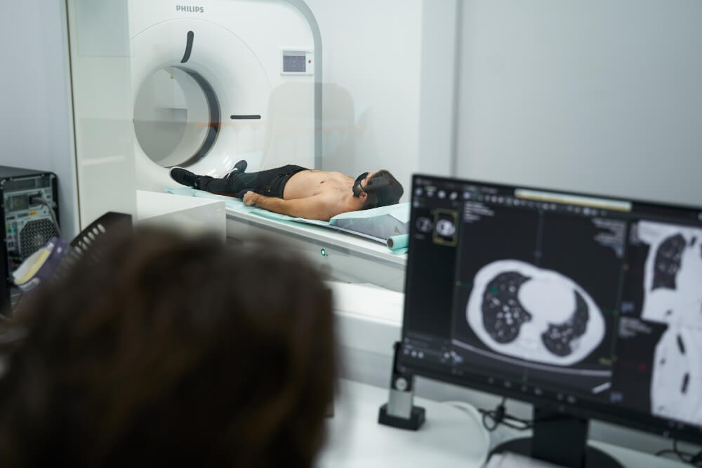
The advancements in CT scanner technology and clinical practice have shown that cardiac CT has numerous advantages over magnetic resonance imaging. Multi-slice computed tomography (MSCT) creates multiple, precise images of the layers in blood vessels, allowing early detection of plaque buildup.
How does coronary artery disease develop?
Coronary artery disease develops when there is damage and/or narrowing of the blood vessels that supply the heart with blood, oxygen, and nutrients. The most common cause of coronary artery disease is the buildup of plaques in the arteries. Deposited plaque narrows the arteries and hinders normal blood flow to the heart. In the early stages, the disease may not present any symptoms, but as plaque accumulates, symptoms such as shortness of breath and chest pain may occur. Complete blockage can lead to a heart attack. Reduced blood flow through the coronary arteries is primarily caused by atherosclerosis of the blood vessels in 95% of cases. Therefore, early detection of coronary artery disease and the degree of artery narrowing, which can be done using CT scanning and magnetic resonance imaging, is crucial.
Advantages of CT scanning over magnetic resonance imaging (MRI)
CT scanning offers several advantages: it is a much faster procedure, typically taking only 10 seconds, compared to MRI which usually lasts at least 30 minutes. Furthermore, CT scanning is suitable for a larger number of patients, as those with pacemakers, implants, or prosthetics are not able to undergo MRI. Another important consideration is that CT scanning is more tolerable for individuals with claustrophobia. With the development of dose reduction software in the past decade, radiation exposure has significantly decreased, resulting in minimal adverse effects from CT scanning.
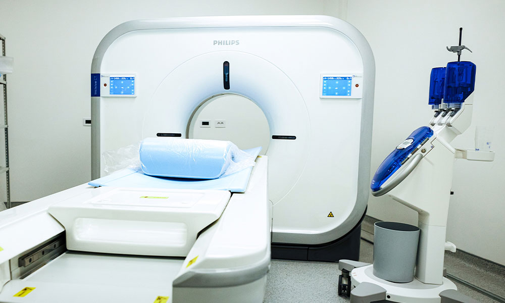
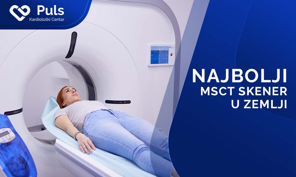
PULS Diagnostic Center possesses a technologically superior, low-dose 128-slice scanner of the latest generation that emits approximately 80% less radiation than other models.
It is true that magnetic resonance imaging (MRI), utilizing a strong magnetic field, provides a detailed depiction of the body part being scanned, in this case, the heart. It is highly sensitive in detecting numerous pathological changes and for planning and monitoring the effects of appropriate therapy. However, computed tomography (CT) scanner (which operates on X-ray radiation) offers an even more precise image of the morphology and overall condition of the specific body part.
Thanks to advanced image post-processing software and artificial intelligence, CT scanning provides a wealth of available information that doctors can use to improve diagnosis, prognosis, and risk assessment not only for the heart but also for other organs.
CT scanning enables the assessment of arterial conditions, not only plaque but also myocardial diseases, as well as other conditions such as lung cancer, osteoporosis, and even breast cancer.
CT scanning provides highly accurate and valuable information for plaque burden classification, performing coronary classification, and assessing functional and morphological changes in the heart. The development of techniques for assessing blood flow and CT fractional flow reserve (FFR) has enabled obtaining both anatomical and functional images in a single scan, with greater efficiency compared to magnetic resonance imaging.
For the majority of patients, cardiac CT is sufficient for making treatment decisions.
CT scanning is also a viable solution for monitoring the condition of postoperative cardiac patients and patients with bypass grafts, as it is safer to assess graft patency using CT coronary angiography rather than conventional coronary angiography.
However, magnetic resonance imaging (MRI) remains a good choice when more information about the myocardium itself is needed.
When is magnetic resonance imaging the preferred method?
MRI scanning takes longer than CT scanning, but it allows for visualization of the function behind stenosis (narrowing) and provides a more comprehensive picture of what is happening in the blood vessel. CT imaging can show stenosis, and software can indicate the degree of stenosis, but it does not show the extent of damage. CT scanning is the best choice for patients without stenosis, such as a 40-year-old woman with atypical chest pain, but it is not ideal when a patient has abnormalities in blood vessels or stenosis. In such cases, CT does not reveal whether they are causing myocardial ischemia.
In conclusion, both CT scanning and magnetic resonance imaging are used to assess the condition of coronary artery disease. Depending on the degree of vascular damage and disease prognosis, the doctor determines whether it is better to perform the diagnostic procedure using a scanner or MRI.