Our kidneys’ main job is to filter our blood. Sometimes we develop masses (growths or tumors) inside our kidneys. Some of these growths are cancerous but many are not. You must have your mass checked out to learn if it is cancerous or not.
There are many different options for treatment. Often there are even more choices if your cancer is found early. Our medical team is there to help you. They can help you learn more about the pros and cons of treatments. Here we share more about kidney cancer and the steps you can take if a mass forms in your body.
Over half of kidney masses are found by chance. Often they are found during generic screening or when you see a doctor about some other problem. If your doctor thinks you may have kidney problems, they might send you to a urologist. A urologist is a doctor who specializes in the urinary system.
There are no routine laboratory tests to find kidney masses. Your health care provider may use many tests to help learn more about your kidneys.
Tests and procedures to exemine kidneys
- Physical exam and history
- Basic or complete metabolic panel (CMP) to check organ function
- Complete blood count (CBC) to check the blood for signs of disease
- Urinalysis to check for infection, blood and protein in urine
- Serum creatinine levels or other kidney function tests to check if the kidneys are getting rid of waste
- Ultrasound to get images of your kidneys
- CT scan and MRI to help diagnose and stage kidney masses
- Bone scan and chest x-ray to find out if the cancer has spread
- Kidney mass biopsy to help find out what type of tumor you have
Symptoms of kidney masses
Most kidney masses have no symptoms in the early stages. If there are symptoms, they will most likely be:
- Hematuria (blood in urine)
- Flank pain between the ribs and hips
- Low back pain on one side (not caused by injury) that does not go away
- Loss of appetite
- Weight loss not caused by dieting
- Fever that is not caused by an infection and does not go away
- Anemia (low red blood cell count)
Diagnosis – What is a Kidney Mass
A tumor, or mass, is an abnormal growth in the body. A kidney mass, or tumor, is an abnormal growth in the kidney. Some kidney masses are benign (not cancerous) and some are malignant (cancerous).
One in four kidney masses are benign. Smaller masses are more likely to be benign. Larger masses are more likely to be cancerous. Some tumors may grow slowly while some can be faster growing – or more aggressive. Aggressive tumors may form, grow and spread very quickly.
Most kidney growths (about 40%) are small, localized masses. Localized means that the tumor has not spread – from where it first started. The main classes of tumors are:
- Renal cell carcinomas (RCC). These are the most common malignant kidney tumors. They are found in main substance of the kidney, where the filtering occurs. RCC may form as a single tumor within a kidney or as two or more tumors in one kidney.
- Benign kidney tumors. About 20% of tumors removed from kidneys are benign. There are about nine named tumors in this class. Some can grow quite large but they are almost always non-cancerous and do not spread to other organs.
- Wilms tumors. Wilms tumors almost always occur in children and are rarely found in adults.
Kidney Embolization
Embolization is a minimally invasive treatment, which is usually used to treat benign (non-cancerous) masses in the kidney. To embolize means to block an artery or vein. During an embolization procedure, small particles are injected through a catheter into a mass. These particles block blood flow to the mass, taking away its supply of oxygen and nutrients. This causes the mass to die and shrink.
In May, 2018, this procedure was performed on ex First Lady Melania Trump to treat a benign kidney mass.
How is the Kidney Embolization Procedure Performed?
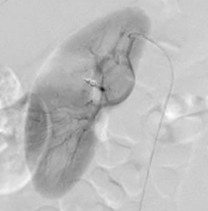
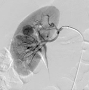
Interventional Radiologist with a team of imaging physicians, highly trained technologists and nurses perform this procedure in a hospital setting. Using X-ray guidance, a specially trained interventional radiologist inserts a thin tube, called a catheter, into an artery in the groin. Once the catheter is positioned in the branches of the artery that are feeding the mass, the embolization particles are injected, specifically targeting the mass. Additional X-rays will be taken to confirm that the mass has been treated. At the end of the procedure, the catheter will be removed and the tiny opening in the skin is then covered with a dressing. No sutures are needed.
A catheter is placed in a kidney lesion
Embolization particles are injected into kidney lesion
Before Your Procedure
- You will be contacted by a member of our team the day before your exam and given instructions on how you should prepare and what time you should arrive. If you are not contacted, please call 0117555000 early in the morning of your procedure
- You should plan to arrive two hours before your scheduled procedure (three hours if you have not had all your pre-op lab work done).
- You should not eat anything from the midnight before your procedure.
- You should consult with your physician about taking your regular medications prior to your exam. Some, such as Coumadin or Plavix, should not be taken before your procedure.
- You will not be allowed to drive after the procedure, so you should arrange for someone to help you get home.
- We want to make your waiting time as pleasant as possible. Consider bringing your favorite magazine, book or music player to help you pass the time.
- Please wear comfortable clothing.
After Arriving
- You will meet with an imaging physician who will explain the procedure to you and answer any questions you might have.
- After this discussion, you will be asked to sign a consent form for the procedure.
- You must notify the nurse, technologist and/or imaging physician of any allergies you may have, or if you are pregnant, prior to your exam.
- A small sample of blood might be drawn for testing.
After Your Procedure
- Some patients will need to stay in the hospital overnight. This will be decided once your procedure is done.
- You should be prepared with a ride home.
- Radiologist will review the images from your exam and write a report of the findings.
- Your doctor will discuss the results with you.
Pulse Cardiology Center provides a full range of advanced imaging, both radiology and cardiology, as well as interventional radiology and interventional tumor treatments.
Grading and Staging of kidney tumors
A tumor grade tells how aggressive the cancer cells are in your body. A tumor stage tells how much the cancer has spread. Grades 1 through 4 show increasing severity with “1” being the lowest level and “4” the highest. A higher grade and more advanced stage usually come with larger tumor size and more aggressive tumors. Tumor size helps in assessing risk for cancer developing.
Kidney cancer is staged using the tumor node metastases (TNM) system.
The “T” in the TNM system tells us the size of the main (primary) tumor and whether it has grown into nearby areas.
T1: Tumor 7.0 cm (about 2 .8 inches) or less, confined to the kidney
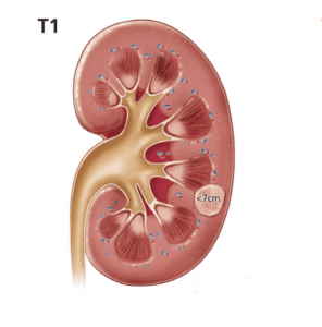
T1a: Tumor 4.0 cm (about 1 .6 inches) or less, confined to the kidney
T1b: Tumor 4.0-7.0 cm, confined to the kidney
T2: Tumor greater than 7.0 cm, confined to kidney
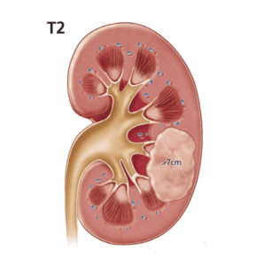
T2a: Tumor greater than 7.0 cm and less than 10 .0 cm, confined to the kidney
T2b: Tumor greater than 10 cm (about 3 .9 inches), confined to the kidney
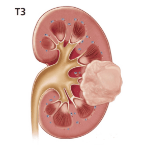
T3: Tumor grows into major veins but not into the adrenal gland and not beyond Gerota’s fascia
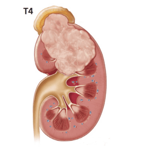
T4: Tumor reaches beyond Gerota’s fascia (including the adrenal gland). This is not a localized tumor.
The “N” in the TNM system tells us how much the tumor has spread to nearby (regional) lymph nodes.
N0: No regional lymph node metastasis
N1: Metastasis in regional lymph node(s)
The “M” in the TNM system tells us about metastasis and whether the cancer has spread (metastasized) to other parts of the body. Spread is most common to the lungs, bones, liver, brain, and far off lymph nodes.
M0: No distant metastasis
M1: Distant metastasis
Stage I and II tumors include cancers of any size that are confined to the kidney.
Stage III tumors are either locally invasive (T3) or involve lymph nodes (N1).
Stage IV tumors have spread beyond the kidney into organs nearby (T4) or distant metastases (M1).