Echocardiography is recommended in many situations. For example, if you belong to the risk group for the development of cardiovascular problems, if you already have symptoms that indicate some problem with a heart, before certain surgeries, or to monitor the course of treatment of already established heart diseases. In addition, even when you don’t have any symptoms or genetic predispositions, a heart echo can be part of your regular annual health check-up.
What Is Echocardiography?
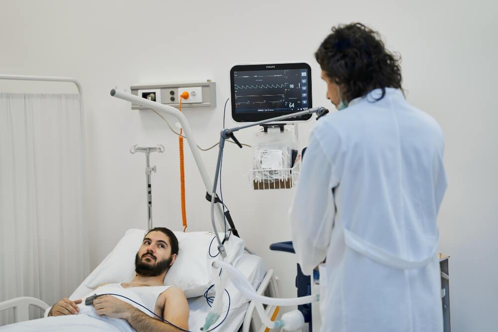
Echocardiography or echo of the heart is a method of imaging the heart that uses two-dimensional, three-dimensional, and Doppler ultrasound to obtain images of the heart in real-time.
In this way, it is possible to see the shape, the size, the performance of the heart, its chambers and atria, the location and dimensions of the damage, whether there is fluid around the heart, murmur, how the heart pumps blood, but also the appearance of the valves.
When Is an Echo of the Heart Necessary?
Echo of the heart can be performed as part of a systematic examination, but also due to the presence of symptoms that indicate the possibility of a heart problem.
If there are specific reasons why ultrasound is recommended, these are usually the following:
- when there is a suspicion of problems with the heart valves or in some cavity of the heart
- when there is a suspicion that the heart tissue is damaged
- if there is a suspicion of a congenital heart anomaly
Preparation for Echocardiography
No special preparation is necessary before the echo of the heart. If you are on any therapy, you should talk to your doctor about whether you should take it regularly or whether it will be useful for you to come to the examination without taking a dose of the medicine.
Types of Echocardiography
In relation to what data regarding the heart are needed by a specialist doctor, the patient can be referred to a certain type of echocardiography. There are the following types:
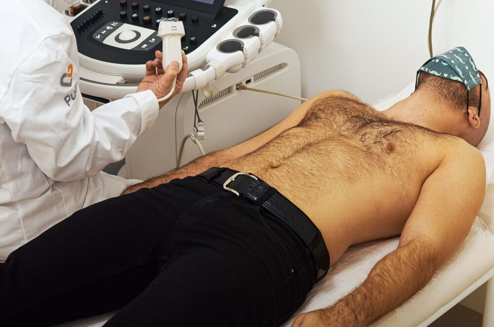
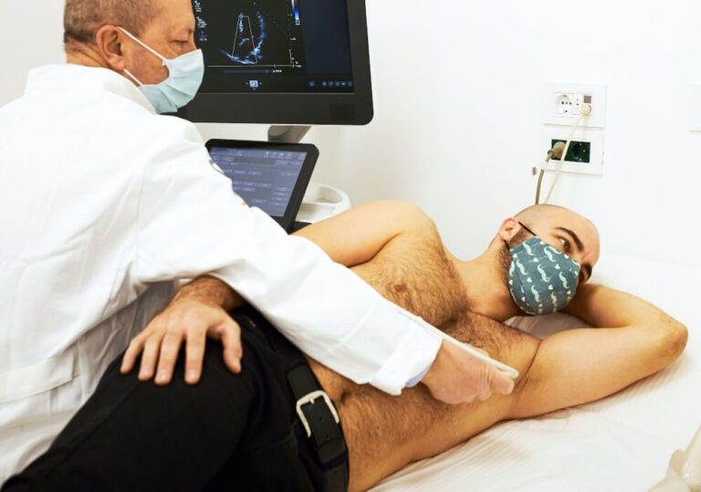
- Transthoracic echocardiography
Transthoracic echocardiography represents a standard ultrasound of the heart. If this type of ultrasound is recommended for you, you should know that there isn’t special preparation for it. During the examination, the patient strips down to the waist, lies on their back, and the doctor applies the gel to the probe and then spreads it on the chest with it. Thanks to the gel, it is possible for ultrasound waves to pass through the chest and visualize the heart.
The doctor will place the probe on the front thoracic wall of the patient, and ultrasound waves will pass through it, creating an image.
During this type of examination, there may be a need to use contrast if the ribs make it difficult to see the heart. Contrast is injected into the patient’s vein. The use of this agent is safe, but it is given only if it is necessary to ensure the quality of the recording.
Besides that, your doctor may ask you to breathe in a certain way or to turn to the left. The examination itself is not unpleasant, except that at some point the pressure of the probe may be stronger in order to better see what the doctor is trying to check.
The ultrasound image can be saved on a hard disk, CD or something similar so that it can be viewed again later.
Echocardiography lasts on average between half an hour and forty-five minutes.
- Transesophageal echocardiography
This method is rarely performed because it is invasive and uncomfortable, but if it is impossible to obtain valid data using standard heart ultrasound, the doctor will resort to this method.
To perform transesophageal echocardiography, a special probe with an ultrasound transducer on the top is needed, which is applied to the patient’s esophagus. In this way, images are obtained directly behind the heart. The obtained recordings have extremely good quality.
It was mentioned earlier that the procedure is unpleasant, so it is recommended that the patient be sedated so that the ultrasound can be done properly.
All the time the doctor will monitor the level of oxygen in the blood, and they may even recommend that the patient stay under observation for some time after the examination, before they are released home.
It is also possible for a person to feel discomfort in the throat after the scan due to the introduction of the probe.
- Doppler echocardiogram
The third type of heart ultrasound is a Doppler echocardiogram. Like the standard heart echo, this is a non-invasive method. Doppler provides more detailed information about the heart and blood vessels. Thanks to this type of examination, it is possible to monitor and measure the speed of blood flow in the heart and check the direction of its movement. The movement of blood is very clearly and vividly depicted because it is colored in two colors: red and blue.
Doppler techniques can be used in transesophageal and transthoracic echocardiography.
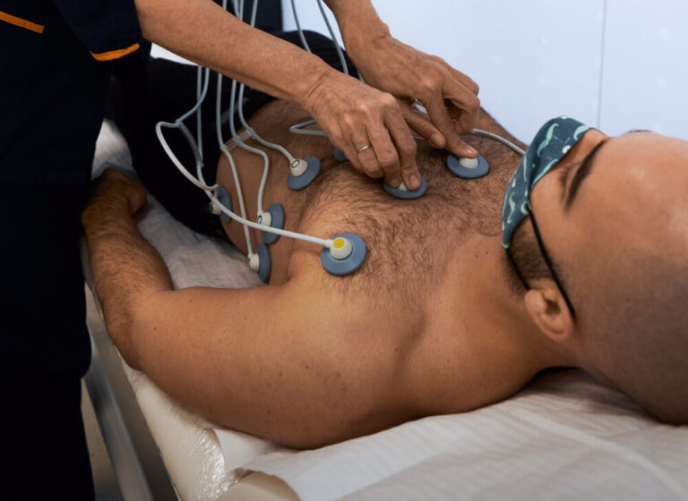
- Stress echocardiography
This procedure is also non-invasive and is used to record the heart wall.
At the very beginning, the doctor takes pictures of the heart at rest, in order to determine the direction of movement of the heart wall.
After that, the patient is instructed to do certain exercises or to walk on the treadmill. The level of load will depend on the age and health condition of the patient. In situations where the patient simply cannot move, the doctor will give medications that will speed up the heart rhythm to the desired or required level.
Then the heart is checked again, but this time “under stress” to monitor the movement of the heart wall at the peak of the heart work.
This ultrasound covers the wall of the heart, not the coronary arteries, but if there are problems with the coronary arteries, they can also affect the movement of the heart wall.
A stress echocardiogram is a relatively safe method. The ultrasound rays that are used are certainly safe, only because of the stress to which the body is exposed, a heart rhythm disorder can rarely occur to a person who has undergone this method, and even less often the consequences can be greater.
- Three-dimensional echocardiography
3D echocardiography is a modern method which, thanks to special probes and data processing systems, gives very detailed views. In this way, it is possible to see the cross-section of the heart in different planes, but also to reconstruct 3D images of anatomical structures, which is important for detecting and understanding pathological processes.
This method has found its application not only in diagnostics but also in some intraoperative procedures, such as endomyocardial biopsy of the right ventricle and the like. It is important in these situations because it can give a real-time picture of the heart.
The Results of Echocardiography
The results of the heart ultrasound will be available to you immediately after the examination is completed. The doctor will talk to you, explain what they have noticed and, if necessary, guide you to further steps. Depending on the results, the person may be referred for additional tests or may be prescribed therapy and explained the course of treatment.
Is Echocardiography a Safe Method?
Ultrasound rays are not harmful, so even children and pregnant women can be exposed to them. There are no limits when it comes to the number of ultrasound examinations, so safety in that sense should not be questioned.
On the other hand, it is important to note that if you do a stress echocardiogram due to the body’s exposure to stress, there is a certain risk, because some side effects can occur in the form of heart disorders, and really rarely induce a heart attack.
People who feel discomfort during exertion, have difficulty with breathing, will probably be referred to this test, to check the characteristics and performance of the heart, its chambers, and the appearance of the valves. It is not necessary to wait long for a call to the doctor and an examination, because in all situations when heart problems occur, a quick reaction is of crucial importance.
Even if you have no symptoms, and for some reason you have a predisposition for developing heart disease, this examination can be useful for you. It is also included in our Total Heart Examination offer, which we formed in order to completely check the hearts of our patients.
With us, you always come first. You will receive an appointment for an examination as soon as possible, and the treatment that awaits you by trained, and professional medical staff will complete the feeling that we are here for you and your health.