X-ray or radiographic imaging is the oldest and most commonly used diagnostic method for visualizing the interior of the body to determine various conditions such as fractures, infections, tumors, lung diseases… The imaging is performed by directing a controlled beam of X-rays from the X-ray tube of the machine towards the part of the body being examined. X-rays pass through the body, and different tissues and structures absorb them in varying amounts, which is reflected in the final image, the radiograph – denser structures such as bones or tumors, which absorb more X-rays, appear brighter, while less dense structures such as soft tissues appear darker. The resulting radiograph provides a two-dimensional representation of the internal body structures, and a radiologist analyzes the image, looks for irregularities, and makes a diagnosis.
When is an X-ray the best solution?
When a doctor will refer a patient for X-ray imaging depends on the specific clinical situation and the problem being investigated, but this method is valuable because it provides quick and relatively inexpensive imaging for many conditions. Here are a few situations in which X-ray is often the best solution:
Skeletal examination
X-ray is the gold standard method for examining bones as it shows fractures, cracks, arthritis, tumors, and other bone and joint abnormalities.
Lung examination
Chest X-ray can detect inflammation, tuberculosis, pulmonary edema, and other lung diseases.
Abdominal examination
Abdominal X-ray can reveal intestinal obstruction, ulcers, inflammatory bowel disease, gallstones or kidney stones, tumors…
Dental examination
Dental X-ray is used to detect cavities, abscesses, and other dental problems.
In addition, X-rays are used in routine screenings such as mammography for breast cancer detection or chest imaging for tuberculosis screening.
How often can one undergo X-rays?
Although X-ray imaging is highly beneficial, exposure to X-rays carries certain risks because this form of ionizing radiation can damage cells in the body and increase the risk of developing malignant diseases, which is why it is applied with caution, especially in pregnant women and children.
However, the risk of such negative effects is very low with occasional routine X-ray examinations. In patients whose clinical presentation requires more frequent imaging or who are more sensitive to radiation, the doctor will consider the balance between the need for diagnostics and potential risks. Other diagnostic methods such as ultrasound and magnetic resonance imaging are also considered to avoid exposure to X-rays.
What is the difference between X-ray and CT scan?
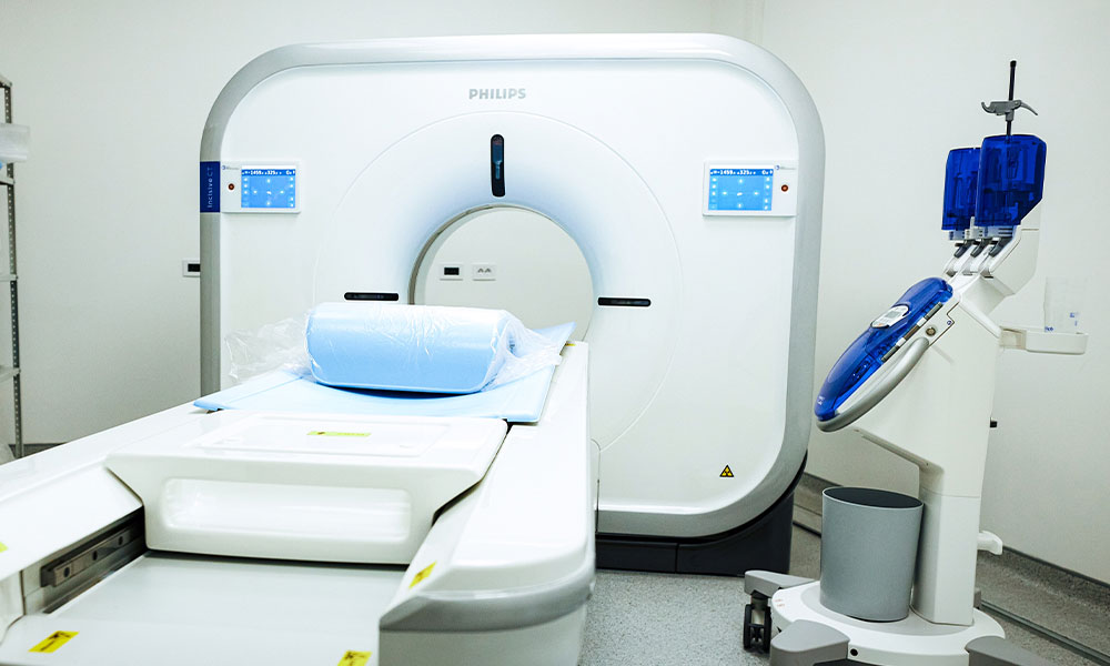
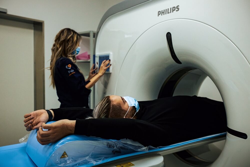
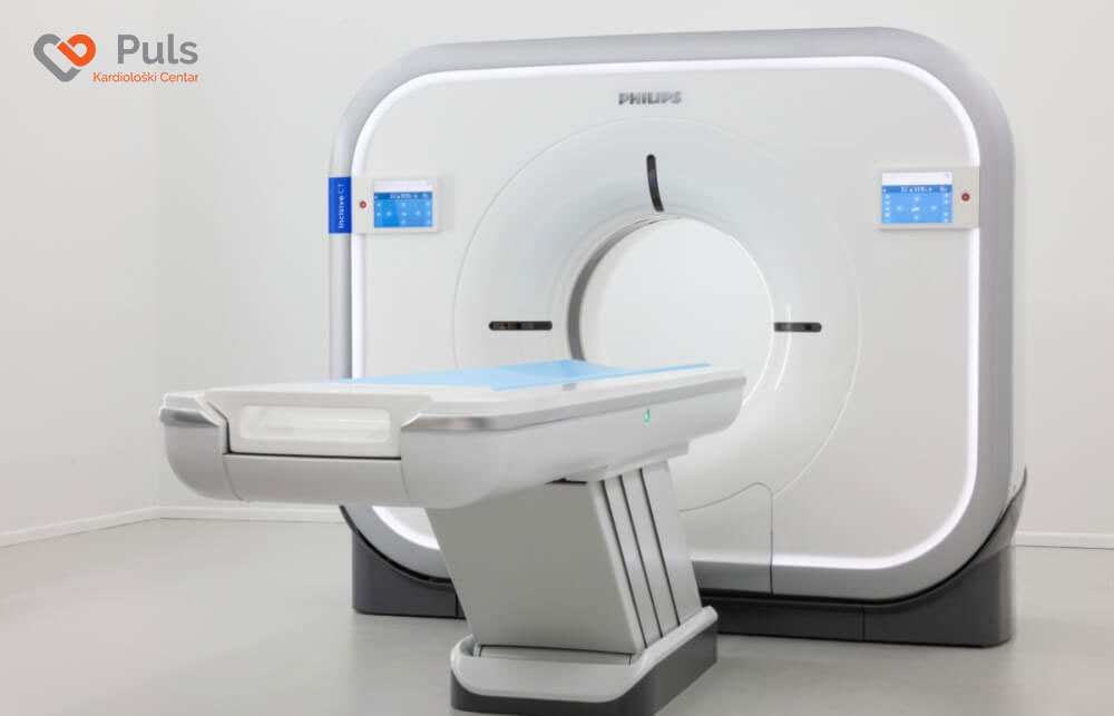
Unlike ultrasound imaging, which is one-dimensional, conventional X-ray provides a two-dimensional image on a conventional X-ray machine and provides important information about the internal structures of the body. With a scanner or computed tomography (CT), although it uses the same X-rays, the image obtained with the help of a computer is three-dimensional and provides more information compared to X-ray or magnetic resonance imaging.
A doctor will, for example, refer you to a scanner if fractures and dislocations of bones need to be determined, then pneumonia or malignant disease. A CT scan will be recommended for more precise diagnostics of internal organ conditions, and sometimes X-ray and CT scans together will help the doctor get the right picture of the condition.
When is an X-ray done for a joint injury?
X-ray imaging of the joint provides the doctor with significant data on the morphological condition of the bone-joint and ligament structures of the joint, and shows changes in the joint that are either the result of traumatic injury with symptoms of pain, swelling, redness, limited movement or degenerative diseases such as osteoarthritis or rheumatoid arthritis.
This diagnostic procedure is most often recommended by an orthopedic surgeon with traumatology specialization, a specialist in physical medicine and rehabilitation, a sports medicine specialist in cases of:
- joint injury such as dislocation
- joint deformity (congenital or acquired, e.g., changes in the fingers of the hands, bunion…)
- swelling in the joint region
- growths in the joint region
- pain in, for example, the knee or hip
- suspicion of inflammation in the joint
- monitoring the postoperative course after joint surgery.
Why can’t medicine do without X-rays?
X-rays were discovered by German physicist Wilhelm Conrad Röntgen on November 8, 1895, quite by accident. Performing an experiment with cathode rays to determine if they pass through glass, he noticed a glow in the dark laboratory even though the cathode tube emitting the rays was covered with thick, black cardboard. In further research, he unsuccessfully attempted to block the rays with various materials, until he finally placed his own hand under it and a photographic plate. He was amazed to see his own bones in the picture projected onto the screen. Six weeks after the discovery, he made the famous first X-ray image of his wife Anna Bertha Ludwig’s hand. For this discovery, he received the Nobel Prize in Physics six years later. His discovery greatly contributed to the development of medicine because it allowed doctors for the first time to look inside the human body without surgery.