Is a CT scan painful?
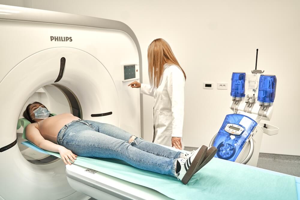
Imaging with a scanner is painless. Scan imaging or MSCT diagnostics is a non-invasive diagnostic method and during the examination you lie on the table while the machine takes the necessary images. If a CT scan is performed with the administration of a contrast agent through a vein, usually in the hand, an iodine-based agent will be injected. When inserting the intravenous cannula, you will feel a slight sting so that the needle is properly placed. After the examination, the cannula will be removed and a small patch will be placed on the puncture site.
How long does a CT scan take?
The examination on the scanner, depending on the region being scanned, can last from 5 to 15 minutes (this includes taking data in the form of a general history, preparation for the examination, which includes an explanation of the examination itself, placement of the i.v. cannula). 90% of CT examinations require the application of iodine contrast agent intravenously, except in the case when bone-joint structures are imaged, which can be scanned without the application of contrast agent.
The CT imaging sequences themselves last from 5 to 8 seconds.
How much radiation does a CT scanner emit?
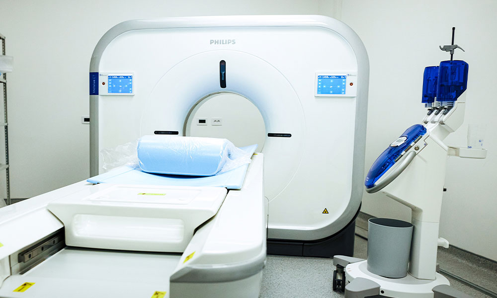
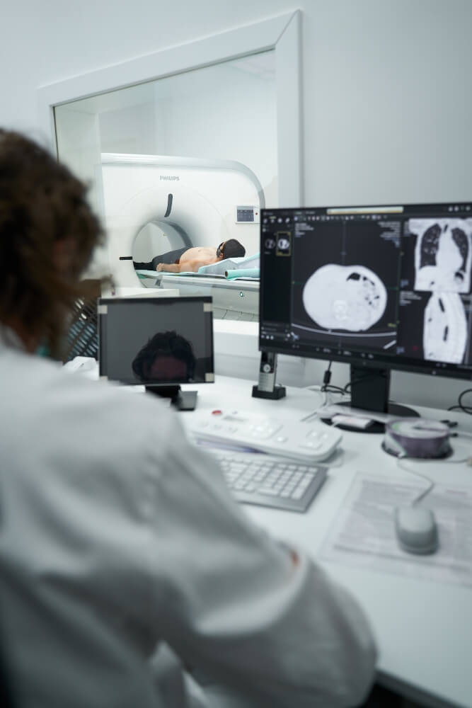
As such, the CT machine works on the principle of an X-ray tube and releases a certain dose of ionizing radiation. X-rays are not retained to such an extent in the human body. The machine we use at Puls Cardiology Center is a state-of-the-art 128-slice Philips Incisive CT scanner. The device has modern software that, according to a given algorithm, calculates the minimum dose of ionizing radiation in order to protect the patient and at the same time provide enough information so that radiologists can interpret the examination and write a report.
For example, a single CT scan of the chest produces 100 to 200 X-rays. That may sound like a lot, but the total amount your body absorbs is still very small.
It is important to know that every day everyone is exposed to ionizing radiation, only that it is radiation from natural radioactive material from the immediate environment. In a year, the average person applies about 3 milliseverts (mSv), the unit scientists use to measure radiation. Each CT scan exposes you to radiation from 1 to 10 mSv, depending on the radiation dose and the part of your body being tested. During a low-dose chest CT scan, you are exposed to radiation of about 1.5 mSv.
Can I get cancer from radiation from a CT scan?
Many people worry when they get a CT scan because radiation is known to be a possible cause of cancer. In fact, the chances of getting cancer from a CT scan are very small. Some organs are more sensitive to radiation than others. Radiation tends to do more damage to rapidly growing and dividing cells. The breast, lungs, thyroid gland and bone marrow have rapidly dividing cells, so they are more sensitive than other parts of the body, such as the brain.
The possibility of developing cancer is slightly higher in women than in men. It is also higher in children, because children grow and their cells divide faster than those of adults.
For many people, a CT scan is worth the minimal risk of radiation exposure compared to the results. A CT scan can help doctors spot dangerous health problems and check if treatment is working. If your medical condition requires several scans in a short period of time, discuss with the radiologist whether the benefits outweigh the risks of radiation. Your doctor will make sure that the benefits you would get from a CT scan outweigh the disadvantages before recommending it.
Overall the chances of getting a fatal cancer from any CT scan are very small.
Allergies to the contrast agent during CT imaging?
The doctor may also want to administer an intravenous (IV) iodine-based contrast agent during the CT scan to highlight blood vessels, organs, and other structures.
If you have an allergy to iodine or have had a reaction to an IV contrast agent in the past, you can still have a CT scan with contrast. Modern IV contrast media are less likely to cause a reaction than older versions of contrast media.
Also, if you are sensitive to iodine, your doctor can pre-prepare you with steroids to reduce the risk of a reaction. If, in addition to everything else, you develop an allergy to the contrast agent, our team of doctors is here, ready to provide all the help and eliminate possible allergic complaints.
Reactions that may occur due to the application of the contrast medium:
Mild reactions: These are relatively common in 3% to 15% of people who receive them. Most of these reactions are mild and include feelings of warmth, nausea and vomiting. Generally, these symptoms are short-lived and do not require treatment.
Moderate reactions: These include vomiting, rash, and swelling, and occur in 0.02% to 2% of people who receive contrast media. They often need treatment.
Severe, life-threatening reactions: These include anaphylaxis, and these reactions occur in 0.04% to 0.02% of people receiving contrast media.
Be sure to discuss and inform your doctor and technician about any allergies you have or questions about potential reactions to the contrast medium.
Do I have an allergy to iodine – iodine contrast?
The question everyone asks before a scheduled CT scan is: “how do I know if I have a contrast allergy”? For now, there is no diagnostic and prognostic test that can reliably predict the risk of adverse reactions to iodine contrast agents. The skin test result is not reliable. A negative skin test does not mean that you will not develop an allergy to iodine contrast medium. What the x-ray technician or radiologist will certainly ask you is information about whether you eat walnuts or nuts or any food that is rich in iodine as a natural compound. If you eat these foods without any allergic reactions, the chance of developing an allergy to the contrast agent during the scan is almost impossible. Every person discovers very quickly during his life whether he is allergic to these foods, since they are very often represented in the diet.
Can I have a CT scan with a pacemaker?
People who have an implanted pacemaker can be subjected to CT imaging without any problems and consequences on the operation of the device and the quality of the imaging itself.
Can I have a CT scan with a defibrillator?
People who have an implanted defibrillator may undergo a CT scan. If the device is placed exactly in the region of the body that is being recorded – that examination will most likely not be suitable for interpretation due to the material from which the device of the same name is made because it can cause disturbances in the image (artifacts). In that situation, classic coronary angiography is recommended instead of CT coronary angiography.
Can I have a CT scan with a hip prosthesis or a knee prosthesis?
One of the advantages of the CT scanner in relation to the magnetic resonance imaging is that the imaging can be done if you have any metal, implanted material or device in your body. People who have any type of hip replacement or knee replacement, who have any metal material in their body, such as bone plates, fixators, or screws, structures, or rods in the spine, can have a CT scan. If you are worried or have any doubts, you can always call us at 011/75-55-000 for all kinds of information.
When will I receive the CT scan report?
The radiological findings of the CT scanner can be obtained within an hour after the imaging at the earliest. Keep in mind that the complexity of the exam and the type of exam can affect the length of time it takes the radiologist to complete an accurate report. In the case when several body segments are recorded, it is possible that you will have to wait longer for your results, and we can certainly send them to your email address if you do not want to wait at the Puls cardiology center. With each report, you will also receive a CD with an image of the scanned body region.
Who performs the imaging on the scanner?
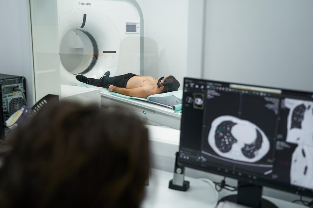
The CT examination itself is performed by a trained X-ray technician. The X-ray technician will collect all the documentation you have brought, talk with you and provide all the information regarding the CT scan itself. When you have received all the answers, the technician will prepare you for the examination. You will be asked to move into the CT diagnostic room and lie on the table, which will move towards the machine that takes the images during the scan. You can communicate with the staff during the recording itself through a speakerphone, and you can communicate visually through the glass that separates the room where the CT machine is located from the control room where the staff sits.

How should I prepare for a CT scan?
Every type of preparation for examination on the CT machine is performed in the premises of our Puls cardiology center. The preparation mainly consists of drinking a certain amount of water, so that the scanned part of the body is better recorded. There are special types of CT scans that require light food on the day of the scan (abdomen, pelvis). You will also tell our team of doctors about all the medicines you are using and any ailments and health conditions you have.
Are any blood tests required before the CT scan?
If a CT examination of soft tissues and blood vessels is indicated (brain and blood vessels, soft tissues of the neck and blood vessels of the neck, lungs, abdomen as a region, pelvis, blood vessels of the legs), it is necessary to perform blood analyses: Urea and Creatinine. If you have the listed results that are not older than 15 days, they will be accepted as valid. If you wish, you can bring ready-made analyzes or we can do them at the Pulse cardiology center. Urea and Creatinine analyzes will be ready within 20 minutes if they are done in our Center.
CT scan or magnetic resonance imaging- MRI
There are several diagnostic methods and examinations that can give us information about the structure of your body, soft tissues, organs, blood vessels.
The biggest difference between a CT scanner and an MRI is that an MRI (magnetic resonance imaging) uses radio waves and a CT scanner (computed tomography) uses X-rays. Most likely, based on your complaints, the doctor will give you a recommendation as to whether you should do an MRI or a CT scan.
If you need a more detailed image of soft tissues, ligaments or organs, your doctor will usually suggest an MRI. Such cases include: herniated discs, damaged ligaments, soft tissues.
If you need a general image of an area such as your internal organs or imaging due to a fracture or head trauma, imaging of blood vessels, a CT scanner is usually recommended.
Differences between CT scanner and MRI
Both MRI and CT can see internal body structures. However, CT imaging is faster and can provide images of tissues, organs and skeletal structures.
MRI is well suited for taking images that help radiologists determine if there are abnormal tissues in the body.
Although both methods are relatively low risk, there are differences that can make each a better option depending on the circumstances.
Risks of a CT scan include:
- possible damage to the fetus due to pregnancy
- a very small dose of radiation
- potential reaction to the use of contrast agents
MRI risks include:
- possible reactions to metals due to magnets
- loud device sounds that cause hearing problems
- rise in body temperature during long MRI scans
- claustrophobia
Your doctor may choose a CT scan instead of an MRI (magnetic resonance imaging) because a CT scan is faster than an MRI scan. In addition, if you are uncomfortable in small spaces, if you are claustrophobic, a CT scanner is definitely a better choice.
An MRI requires you to be in an enclosed space while loud noises are heard around you. In addition, an MRI is much more expensive than a CT scan.
Your doctor may choose a CT scan instead of an X-ray because the scanner provides more detail than an X-ray. The CT scanner rotates around your body and takes pictures from different angles. X-rays only take pictures from one angle.